The New Truth About Brain Immunity — Did You Know? | A Look at the Recent Science Review
Release time:2025-04-09 14:58:15
Literature Interpretation
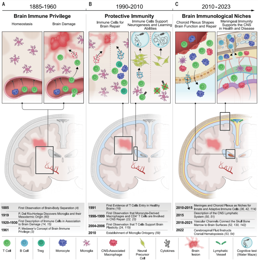
In the past, scientists believed the brain was "immune privileged"—separated from the immune system by an insurmountable barrier. In 1885, researchers observed that when dye was injected into the periphery of animals, it could not enter the brain. This phenomenon reinforced the belief that the brain and immune system operated independently. Furthermore, brain tissue transplants elicited weaker rejection responses compared to peripheral tissues, strengthening the idea of brain immune privilege. Even after the discovery of the brain's lymphatic system, many still held onto this view, assuming that the lack of traditional lymphatic circulation meant the brain was isolated from the immune system. However, with continued research, this traditional notion has been completely overturned. We now understand that the immune system plays a critical role in brain development, daily function, and repair processes. Immune cells, like loyal guardians of the brain, are distributed throughout key brain regions, safeguarding its health. The review article "Transforming the understanding of brain immunity", written by Giulia Castellani and colleagues and published in Science, summarizes the major advances in brain immunity research. It outlines the shift in perception from “immune privilege” to “close integration” between the brain and the immune system, explores brain-resident immune cells, the brain’s immune microenvironment, and related disease mechanisms, and provides insights into therapeutic strategies—offering an important reference for understanding brain immunity and developing treatments for related disorders.
Since the 20th century, studies have revealed the involvement of the immune system in autoimmune inflammatory diseases and neurodevelopmental disorders, leading to the proposal of the concept of "protective autoimmunity." In addition, researchers have identified immune microenvironments within the brain—including the meninges, choroid plexus, and perivascular spaces—as well as lymphatic drainage pathways and connections to skull bone marrow. These findings suggest that the brain is not completely isolated from the immune system but is instead engaged in intricate interactions with it. This shift in understanding has laid a critical foundation for subsequent research.
Figure 1 illustrates a timeline of major milestones in the evolving understanding of brain immunity. Key advances that transformed our perception of brain-immune interactions can be categorized into three periods:(A) Discoveries in the first half of the 20th century supported the concept of brain “immune privilege,” which posited that immune cell infiltration was always associated with inflammation or disease states in the central nervous system (CNS).(B) Beginning in the late 20th century, early findings started to reveal the roles of immune responses in maintaining and repairing CNS homeostasis, and later, in influencing neurogenesis and cognition.(C) The identification of distinct immune microenvironments within the brain showed that both innate and adaptive immune cells residing in or trafficking through these compartments can regulate brain function and repair processes.
2. Immune Cells in the Brain
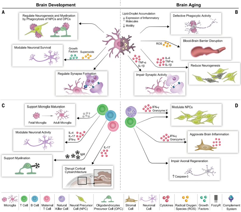
Myeloid Cells:Among myeloid cells, microglia begin functioning as early as the embryonic stage. Starting from embryonic day 9.5, microglial precursors migrate from the yolk sac into the developing central nervous system. These cells are not only involved in regulating neurogenesis and neuronal survival but also contribute to the process of myelination. In adulthood, microglia play a vigilant role in maintaining brain homeostasis and are often referred to as the brain’s “patrolling sentinels.” However, as the brain ages, microglia may become “overwhelmed” and less effective. In the aging brains of both humans and mice, microglia accumulate lipid droplets, increase the production of reactive oxygen species and pro-inflammatory cytokines, and show diminished phagocytic ability. These changes can disrupt the blood–brain barrier and negatively affect adult neurogenesis and synaptic function, closely linking microglial dysfunction to the pathogenesis of neurodegenerative diseases such as Alzheimer’s and Parkinson’s. Adaptive Immune Cells:In addition to myeloid cells, adaptive immune cells also play indispensable roles in the brain. While it remains unclear when lymphocytes begin infiltrating the central nervous system, studies have shown that depleting CD4⁺ T cells in mice at postnatal day 5 impairs microglial maturation and leads to a variety of behavioral abnormalities. In adulthood, CD4⁺ T cells are critical for maintaining normal brain function—they help the brain cope with psychological stress, support cognitive function, and maintain healthy social behavior. However, with aging, the immune cell balance within the brain is disrupted. For instance, CD8⁺ T cells secreting interferon-gamma (IFN-γ) infiltrate neurogenic regions in aged mice and suppress neural stem cell proliferation. Similarly, the accumulation of natural killer (NK) cells also contributes to the decline in neurogenesis.
Figure 2 illustrates the roles of microglia and adaptive immune cells in brain development and aging:(A) Mechanisms by which microglia support healthy brain development;(B) Morphological and functional changes in aged microglia and their impact on age-related neurodegeneration;(C) The roles of B and T lymphocytes in brain development;(D) Mechanisms by which lymphocyte infiltration into brain parenchyma promotes brain aging.
3. The Immune Microenvironment at the Borders of the Central Nervous System
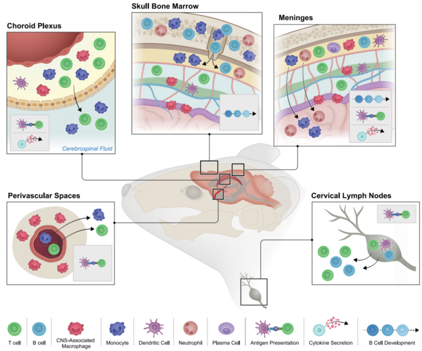
Meninges:Once regarded merely as a “shield” protecting the brain, the meninges are now recognized as an immunologically active “battleground” teeming with immune cells. This region contains lymphocytes and antigen-presenting cells capable of presenting antigens and regulating immune cell recruitment and homing. For instance, γδ T cells in the meninges can modulate anxiety-like behavior by influencing glutamate-releasing neurons in the cortex. Additionally, IgA⁺ plasma cells originating from the gut act as loyal “guardians,” stationed near the dural venous sinuses to prevent pathogens in the bloodstream from invading the brain. Choroid Plexus:Located within the brain ventricles, the choroid plexus not only regulates the composition and volume of cerebrospinal fluid (CSF) but also serves as a critical “gateway” for immune cells entering the brain. Macrophages in the choroid plexus exist in distinct subtypes and can sense and respond to peripheral inflammation. In conditions such as acute CNS injury or amyotrophic lateral sclerosis (ALS), the choroid plexus becomes a key entry point for T cells and macrophages. However, with aging or in neurodegenerative diseases, IFN signaling in the choroid plexus becomes dysregulated, negatively affecting brain function. Perivascular Spaces:The perivascular spaces act as the brain’s “waste clearance stations,” surrounding cerebral blood vessels and hosting immune cells that help regulate CSF flow and clear antigens, metabolic byproducts, and other molecules from the central nervous system. In neurodegenerative diseases, activation of perivascular cell adhesion molecules (CAMs) has been shown to reduce levels of pathogenic amyloid-β42, offering new therapeutic insights for related disorders.
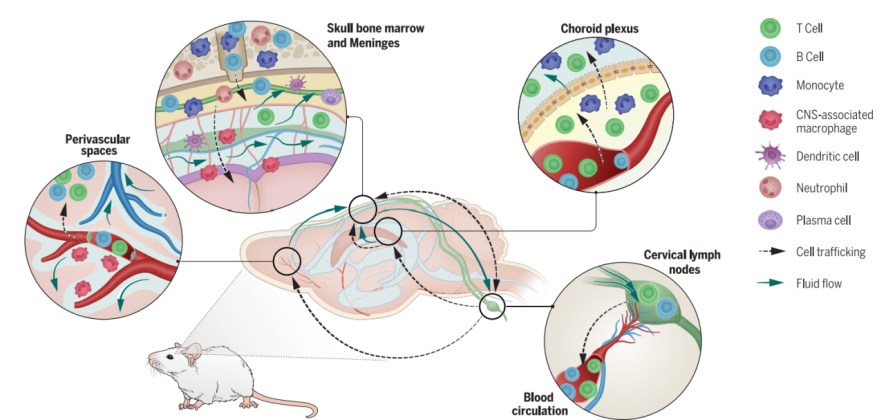
Figure 3. Brain Borders and the Pathways Connecting the Periphery to the Brain. This figure illustrates the distribution of immune cells within and around the mouse brain, highlighting the types of cells that home to distinct immune microenvironments under steady-state conditions. These compartments include the perivascular spaces, choroid plexus, skull bone marrow, meninges, and draining cervical lymph nodes. The figure also depicts immune processes occurring in these regions under both physiological and pathological conditions, such as antigen presentation, cytokine secretion, and B cell development. Arrows indicate the major pathways—confirmed under various disease states—through which these immune niches communicate with the brain.
4. Immune Regions Connected to the Central Nervous System
Skull Bone Marrow (Skull BM):Research has shown that immune cells from the skull bone marrow can directly migrate to the meninges, and CSF can influence the microenvironment and hematopoiesis within the skull bone marrow. In disease states such as multiple sclerosis, changes in immune cells in the skull bone marrow may participate in disease progression. The skull bone marrow is a crucial component of the brain's immune system, with close cellular and fluid communication between the brain and the marrow. This suggests that it may play a key role in brain immune responses and disease development. Cervical Lymph Nodes (CLNs):The cervical lymph nodes drain extracellular fluid from the brain through the meningeal lymphatic system and serve as a site where peripheral lymphocytes encounter brain antigens, potentially triggering adaptive immune responses. During aging and in various disease conditions, changes in the function of cervical lymph nodes can impact the brain's immune status and cognitive abilities. Studies have shown that removal or ligation of cervical lymph nodes in mice leads to cognitive decline, emphasizing the critical protective role of the CLNs in maintaining brain health.
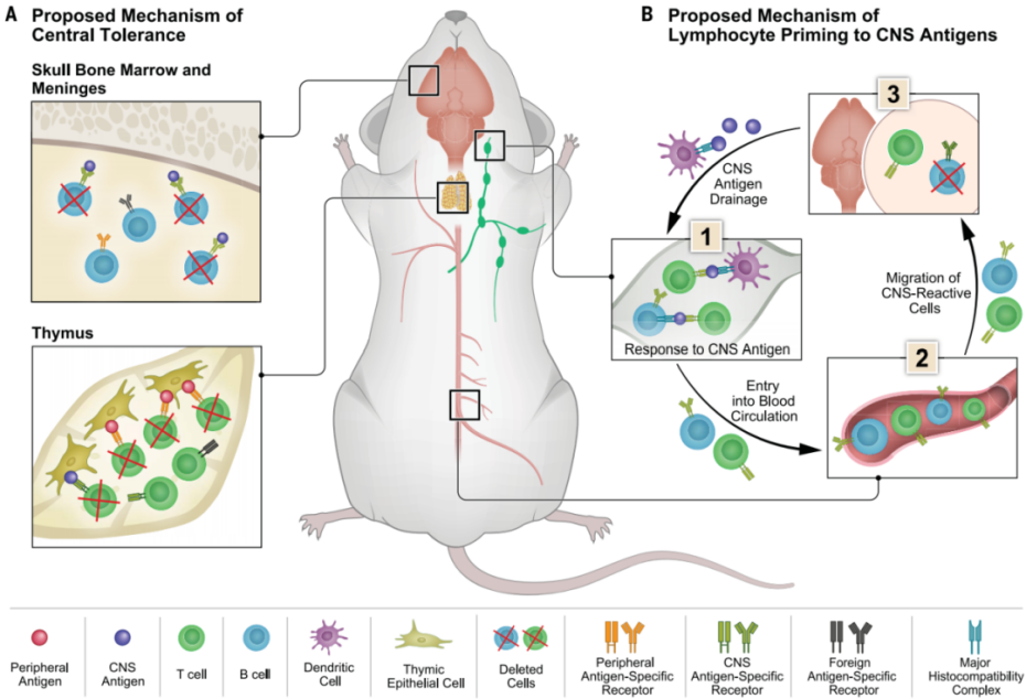
Figure 4 illustrates mechanisms of immune tolerance induction, lymphocyte differentiation, and the homing of CNS-reactive immune cells to brain borders:(A) B cells originating from the Skull BM are cleared in the meninges, while potentially autoreactive T cells with high affinity in the thymus are also eliminated through biological processes. Although high-affinity autoreactive T cells are excluded, low-affinity autoreactive T lymphocytes may escape the thymus.(B) This model explains a series of events following CNS antigen drainage to the CLNs, where they are presented to lymphocytes (1); after immune responses are triggered (2), CNS-reactive lymphocytes enter the bloodstream and eventually home to the CNS borders (3).
5. A New Understanding of Brain Immune Privilege
The concept of brain immune privilege has been revisited in light of new research findings that challenge traditional views. The brain relies on the integrity of the immune system, with a complex immune network in place that involves the recruitment of immune cells and the regulation of immune microenvironments. Rather than being an immune isolation, the brain’s immune privilege involves the precise regulation of immune cell participation. The concept of brain immune privilege needs to be redefined, as there is a complex and finely tuned interaction between the brain and the immune system. This understanding provides a theoretical basis for developing immune-based therapeutic strategies for brain diseases.
Figure 5 illustrates the new understanding of brain immunity. This figure demonstrates a communication network that enables precise regulation of brain immune surveillance. Immune cells are strategically distributed within immune microenvironments, where they are in continuous contact with central nervous system signals transported by intracerebral interstitial fluid, cerebrospinal fluid, and lymph. Upon detecting signals from the brain, immune cells can migrate toward the central nervous system to exert their effector functions.
6. The Link Between Brain Immunity, Disease, and Treatment
Imbalance in brain immunity is associated with a variety of conditions, including aging, autoimmune diseases, neurodegenerative diseases, and neurodevelopmental disorders. For instance, in diseases such as Alzheimer's disease and Parkinson's disease, abnormal changes in immune cells contribute to disease progression. Based on the new understanding of brain immunity, there is hope for developing novel therapeutic strategies for these diseases, such as regulating immune cell functions and improving the immune microenvironment. Brain immune imbalance plays a significant role in various diseases, and a deeper understanding of brain immune mechanisms could provide new targets and strategies for disease treatment.