Tumor Models - Animal Tumor Models - Model Tumor - BrainCase ()
Several common animal tumor models
In order to evaluate the effects of cancer therapy in preclinical and clinical studies, the establishment of ideal tumor animal models provides a good alternative to clinical trials and is an important part of anti-tumor research. The establishment of tumor animal model can evaluate the efficacy of anti-tumor immunotherapy, serve as a screening model for anti-tumor drugs, provide a powerful tool for the study of tumor pathogenesis and metastasis, and enable researchers to understand the mechanism of clinical drug action, which plays an important role in the research and development of oncology and anti-tumor drugs. With the deepening of the understanding of tumor and the development of experimental zoology, as well as the application of some new techniques, the study of tumor animal models has also made important progress. The establishment of tumor animal model can be used to: (1) evaluate the efficacy of anti-tumor immunotherapy; (2) As a screening model for anti-tumor drugs; (3) To provide a better research platform for tumor metastasis research; (4) To provide a good experimental tool for the development of anti-metastatic drugs. Braincase Biotechnology Co., Ltd. provides: human gastric cancer, human liver cancer, human lung cancer, breast cancer, pancreatic cancer, prostate cancer and other human or mouse tumor cell tumorigenesis experiment, and related drug sensitivity analysis experiment; To provide customers with tumor in situ implantation, tumor metastasis model construction, induced tumor model construction, drug sensitivity analysis and other related services.
Classification of tumor animal models
According to the transplantation method of tumor animal model, tumor model can be divided into subcutaneous transplantation tumor, subcapsular transplantation tumor, orthotopic transplantation tumor and blood transplantation tumor.
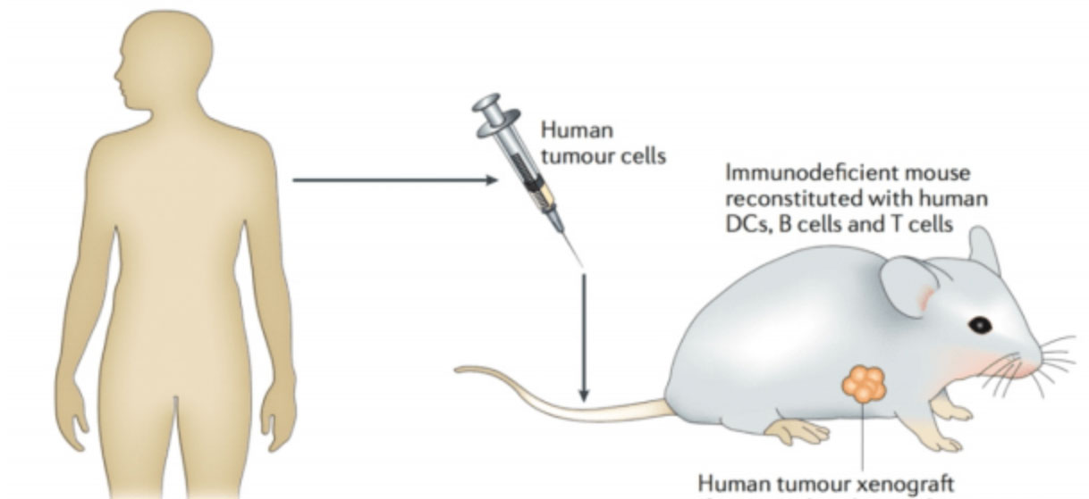
- 1.Subcutaneous tumor transplantation model
As the name suggests, this model is built by planting tumor cells or tumor tissue directly under the skin of mice. The point of implantation is also particular, and the groin and armpit are generally selected for rich lymphatic return. The transplantation site can be selected according to the experimental design, and the location of the transplantation site can be unified. In addition to complying with the unified conditions of the experiment, it is also beautiful to leave photographic evidence when the tumor is collected after maturity. Nude mice (Balb/c mice, hairless mice, T lymphocyte deficiency) are the most common and commonly used experimental mice, especially in the establishment of subcutaneous transplanted tumor tumor models. The establishment of transplanted tumor model in nude mice has the advantages of short establishment period, high tumor formation rate, easy operation and low cost. Of course, this tumor model also has a defect, that is, it cannot accurately simulate the development of normal human tumors.
- 2.Renal subcapsular transplantation tumor model
Renal subcapsular blood flow is more abundant, which is more suitable for those tumor cells that require harsh nutritional environment. Mice used for renal subcapsular tumor transplantation are mainly NOD-SCID mice (hybrid SCID mice and nonobese diabetic mice (NOD) with defects in T and B lymphocytes, antigen presenting cells, circulating components and some NK cells). The phenotype of these mice is white haired mice. The feeding conditions are harsh, and a sterile SPF feeding environment is required.
- 3.Orthotopic transplantation tumor model
In order to better simulate the growth of the tumor, the organ with tumor was selected as the transplantation site. The commonly used mice are still NOD-SCID mice. Open surgery was performed to open the corresponding mouse organs exposed to the corresponding tumors, and tumor tissue or tumor cells were implanted into the organs. This method is not often used because it is not easy to operate and the mortality rate of mice after operation is high.
- 4.Blood transplantation tumor
The common hematologic tumors include leukemia, multiple myeloma (MM) and lymphoma, and the corresponding mouse models can be established for drug-resistant relapsed and refractory clinical patients.
Product Directory
neoplasms
histologic type |
cell line |
scope of application |
breast cancer |
MDA-MB-231 |
Balb/c nude、C-NKG/in situ |
colon cancer |
Colo205, HT29 |
Balb/c nude、C-NKG/subcutaneous |
HCT116, SW480 |
Balb/c nude、NOD-Scid、C-NKG/subcutaneous |
SW620 |
NOD-Scid、C-NKG/subcutaneous |
lung cancer |
NCI-H1299, NCI-H1975, NCI-H460 |
Balb/c nude、C-NKG/subcutaneous |
PC-9 |
SCID-Beige、C-NKG/subcutaneous |
HCC827 |
Balb/c nude、C-NKG/subcutaneous |
A549 |
Balb/c nude、NOD-Scid、C-NKG/subcutaneous |
lymphoma |
Raji |
NOD-Scid、C-NKG/subcutaneous,caudal vein |
melanoma |
A375 |
Balb/c nude、C-NKG/subcutaneous |
ovarian cancer |
OVCAR3, SK-OV-3 |
Balb/c nude、NOD-Scid、C-NKG/subcutaneous |
pancreatic cancer |
PanC-1 |
Balb/c nude、C-NKG/subcutaneous |
AsPC-1, BxPC-3 |
Balb/c nude、NOD-Scid、C-NKG/subcutaneous |
gastric cancer |
HGC-27 |
Balb/c nude、NOD-Scid、C-NKG/subcutaneous |
liver cancer |
Hep G2 |
Balb/c nude、NOD-Scid、C-NKG/subcutaneous |
HuH7 |
NOD-Scid、C-NKG/subcutaneous |
NCI-H1299 |
Balb/c nude、C-NKG/subcutaneous |
cervical cancer |
Hela |
Balb/c nude、NOD-Scid、C-NKG/subcutaneous |
prostatic cancer |
22RV1 |
NOD-Scid、C-NKG/subcutaneous |
Brain Case has SPF-level animal facility qualifications and extensive experience in animal modeling and evaluation, which maximizes the success rate of modeling. If you are interested in the details of the experiment or the problems that may arise during the experiment and their causes, please contact: bd@braincase.cn
literature citation
- Alteri, E.; Guizzaro, L. Be open about drug failures to speed up research. Nature 2018, 563, 317–319.
- DiMasi, J.A.; Reichert, J.M.; Feldman, L.; Malins, A. Clinical approval success rates for investigational cancer drugs. Clin. Pharmacol. Ther. 2013, 94, 329–335.
- Ledford, H. Us cancer institute overhauls cell lines. Veteran cells to be replaced by human tumours grown in mice. Nature 2016, 530, 391.
- Mestas, J.; Hughes, C.C. Of mice and not men: Differences between mouse and human immunology. J. Immunol. 2004, 172, 2731–2738.
- Kojima, Y.; Hayakawa, F.; Morishita, T.; Sugimoto, K.; Minamikawa, Y.; Iwase, M.; Yamamoto, H.; Hirano, D.; Imoto, N.; Shimada, K.; et al. Ym155 induces apoptosis through proteasome‐dependent degradation of mcl‐1 in primary effusion lymphoma. Pharmacol. Res. 2017, 120, 242–251. 6. Flanagan, S.P. ʹNudeʹ, a new hairless gene with pleiotropic effects in the mouse. Genet Res. 1966, 8, 295–309.
- Budzynski, W.; Radzikowski, C. Cytotoxic cells in immunodeficient athymic mice. Immunopharmacol. Immunotoxicol. 1994, 16, 319–346.
- Giovanella, B.C.; Fogh, J. The nude mouse in cancer research. Adv. Cancer Res. 1985, 44, 69–120.