Optogenetics - A Nobel-level tool in neuroscience research
Optogenetics refers to technology that combines optical and genetic means to precisely control the activity of specific neurons. This technology uses genetic manipulation techniques to introduce exogenous light-sensitive protein genes into living cells and express light-sensitive channel proteins on the cell membrane structure. Then, through the irradiation of light of a specific wavelength, the activation and closing of the light-sensitive channel protein on the cell membrane structure is completed to control the opening and closing of the ion channel, thereby changing the changes in the cell membrane voltage, such as membrane depolarization and hyperpolarization. When membrane voltage depolarization exceeds a certain threshold, neurons will be induced to produce transmissible electrical signals, that is, neuron activation. On the contrary, when the membrane voltage is hyperpolarized to a certain level, the generation of neuronal action potentials will be inhibited, that is, neuronal inhibition.
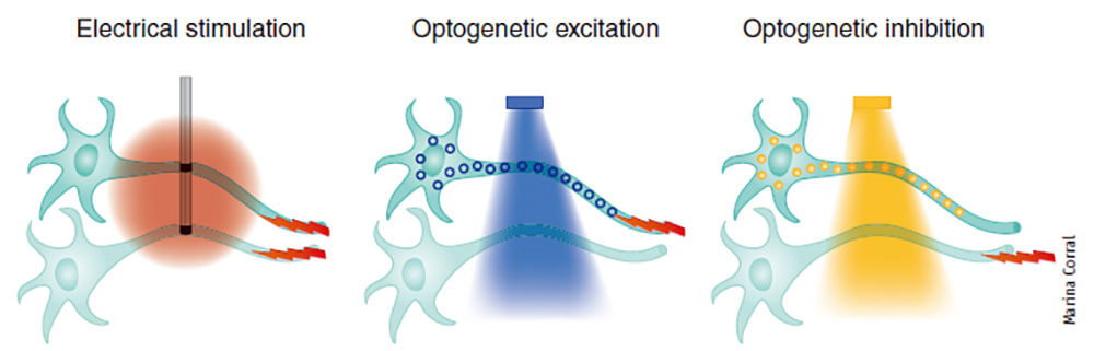
Traditional electrophysiology activates multiple types of neurons with high temporal resolution. Compared with electrophysiology, optogenetic technology can control the activity of specific neurons without or with low damage to study the function of the neural network. It is especially suitable for in vivo and even awake animal behavioral experiments, with high spatiotemporal resolution and high specificity and axonal projection selectivity (Figure 1).
Figure 1. Differences between optogenetics and electrophysiological regulation of cells (KarlDeisseroth, et al., Annu.Rev.Biomed.Eng., 2014)
The principle of optogenetic technology
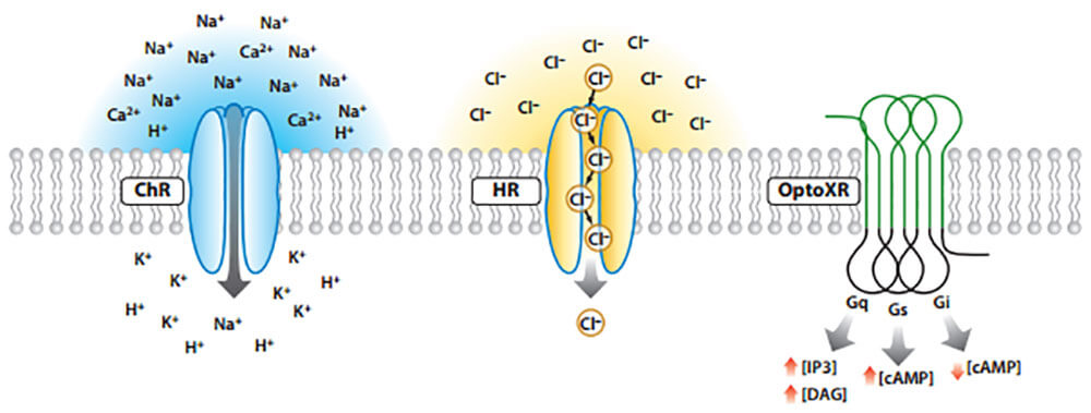
The activity of cells regulated by optogenetic technology depends on the type of light-sensitive channel proteins, namely excitatory light-sensitive channels and inhibitory light-sensitive channels. If the channel transferred into the cell is ChR, when the cell is irradiated with blue light, the channel opens and a large amount of cations flow in, generating a depolarization-evoked action potential and activating the cell. If the channel transferred into the cell is HR, when the cell is irradiated by yellow light, the channel will open, and a large amount of anions will flow in, resulting in hyperpolarization, making it difficult for action potentials to be sent out, and inhibiting cell activity. In addition, there is a type of light-activated or inhibited channel, optoXR. After being activated by light of a certain frequency, it changes the intracellular kinase system and affects cell activities (Figure 2).
Figure 2. Types of optogenetic technology that regulates cell activity (Karl Deisseroth, et al., Annu.Rev.Neurosci, 2011)
Three commonly used optogenetic techniques
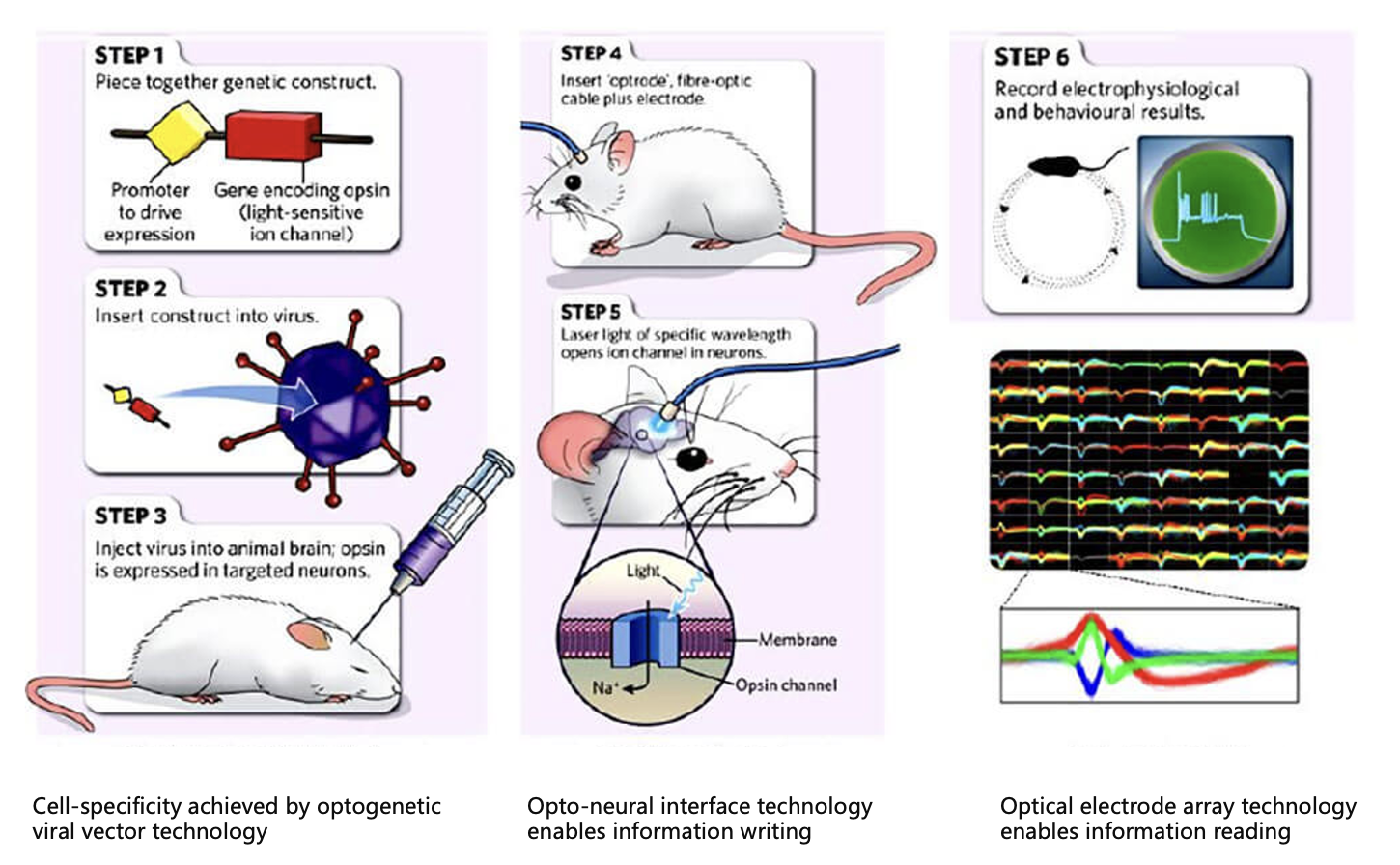
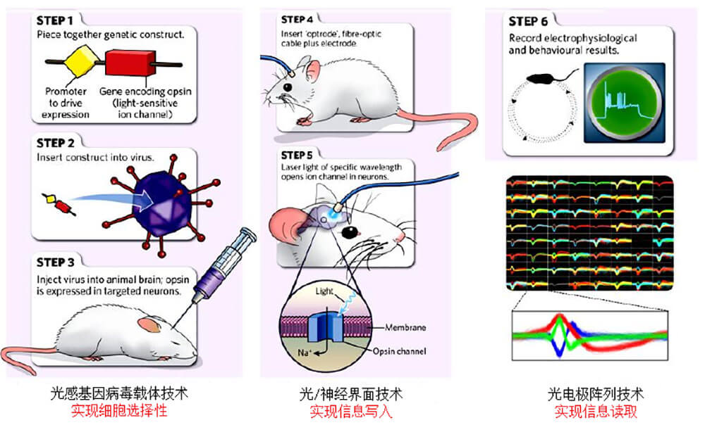
I Optogenetic electrophysiology(The experimental steps are as follows:):
1) Select the appropriate light-sensitive channel protein according to the needs of the experimental design;
2) Insert the appropriate light-sensitive channel into the viral vector and infect the cells so that the light-sensitive channel protein is expressed in specific types of cells;
3) Implant optical fibers into the brains of experimental animals;
4) According to the expression time of the virus, the development of light-sensitive channels is controlled by laser with specific frequency;
5) Record experimental results through electrophysiology.
Application : In vivo circuit functional verification.
Figure 3. Experimental steps of optogenetics (BuchenL, Nature, 2010)
II. Optogenetic patch clamp (Upstream optogenetic combined with downstream patch clamp)(The experimental steps are as follows:)
1) Select mice of appropriate age (generally 6-7w) according to experimental needs, and stereotaxically inject AAV carrying the light-sensitive protein into the upstream brain area A (the virus volume needs to be adjusted according to the virus titer and nuclear mass size).
2) After the virus has been expressed for 2-3 weeks, decapitate the mouse, take out the brain, and slice it into sections with a thickness of 300 μm (target brain area A).
3) Select cells in good condition that are fluorescently labeled by AAV for recording, use single and different frequency lasers to stimulate the target brain area A, and observe and record the response of the cells. If there is a response, it is confirmed that the virus is working and the cells are in good condition, which can further determine the follow-up. Stimulation frequency required for the experiment; if there is no response, the cell status needs to be determined first. If the cell status is good, the stimulation intensity can be increased to determine whether the virus is working. After confirming that the labeled cells in brain area A respond to light stimulation, subsequent experiments can be conducted.
4) After the virus has been expressed for at least 3 weeks, the mice are decapitated, the brains are taken out, and the slices are sectioned by shaking, with a thickness of 300 μm (target brain area B).
5) Select the cells in good condition near the nerve fibers labeled by AAV fluorescence in brain area B for recording, stimulate the target brain area B with light, and observe the response of the recorded cells. If there is a response, observe the relationship between the cell response and the light stimulation. time interval to determine whether it is a monosynaptic connection; if there is no response, the cell status needs to be determined first. If the cell status is good, the stimulation intensity can be increased and then recorded. After confirming that the cells in brain area B respond to light stimulation, subsequent experiments can be conducted.
6) By adding agonists or antagonists of different receptors to the recording tank, the type of synaptic connection can be further determined.
7) Repeat the above steps 5 and 6 for different cells and different brain slices in the same brain area, obtain a certain amount of data and then analyze, organize and draw graphs.
Application: In vitro circuit function validation; determining functional connectivity between two brain regions (upstream region A and downstream region B).
III. Optogenetic behavioral science(The experimental steps are as follows:)
1) Find suitable opsins that serve as reporter genes, including BR, HR, ChR, CoChR, etc.;
2) Deliver light-sensitive proteins to recipient cells to complete transfection and expression;
3) Select appropriate light sources for regulation, and accurately regulate the corresponding biological reactions of cells, tissues and organs by changing the wavelength of light and the intensity of light pulses;
4) Observe animal behavior after light stimulation and evaluate the effects.
Application : Animal behavior research (including eating behavior, reward behavior, anxiety and depression behavior, pain behavior, etc).
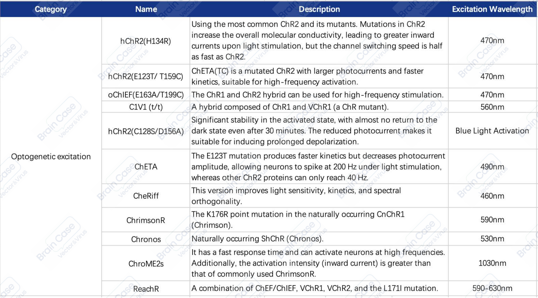
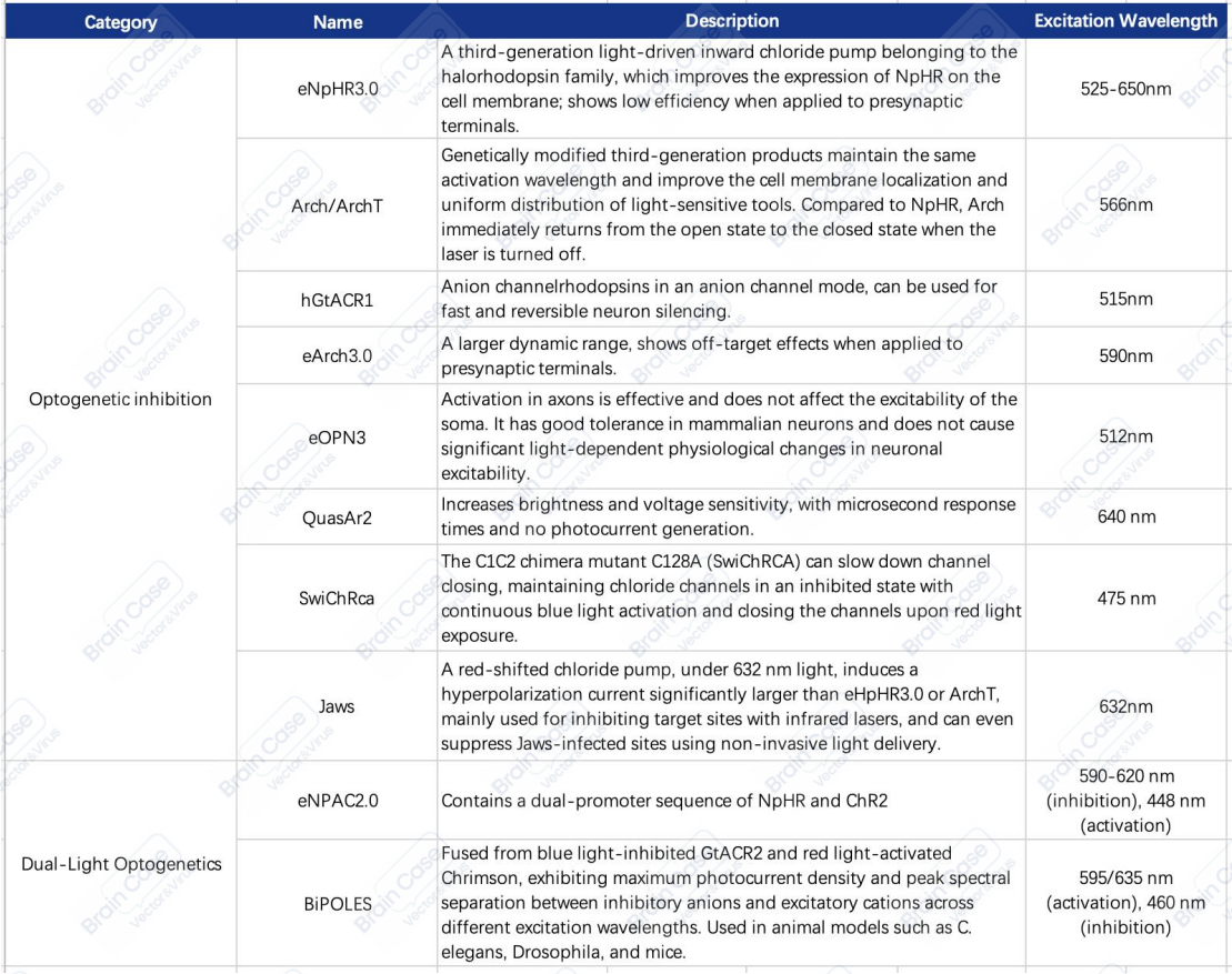
Common photosensitive channel proteins and their characteristics are shown in the table below:
Table 1. List of Optogenetic excitation
Table 2. List of Optogenetic inhibition and Dual-Light Optogenetics
Brain Case offers experimental services including optogenetic and chemogenetic manipulation, fiber photometry for calcium and neurotransmitter signal recording, EEG, EMG, and patch-clamp electrophysiology. These services are well-suited for studies of neural activity and function, as well as for evaluating neuropharmacological and neurotoxicological effects.
If you are interested in the details of the experiment or possible problems and causes during the experiment, please contact: BD@ebraincase.com
Service Type :
Select the service you'd like to purchase.
Order Information(Premade-AAVs)
Please provide us some information about the service you'd like to order.
Order Information(Custom AAV/Lentivirus)
Please provide us some information about the service you'd like to order.
Order Information(Others)
Please provide us some information about the service you'd like to order.