World Parkinson’s Day Special: Microglia in Focus – From Inflammation to Intervention in Parkinson’s Disease
Release time:2025-04-11 13:43:42
Parkinson’s disease (PD) is a common neurodegenerative disorder of the central nervous system, ranking as the second most prevalent form of progressive dementia after Alzheimer’s disease. It is estimated to affect approximately 10 million people worldwide, with a higher incidence among the elderly and an average onset age of around 60 years. The hallmark pathological feature of PD is the progressive degeneration and loss of dopaminergic (DA) neurons in the substantia nigra of the midbrain, which leads to a significant reduction in DA levels in the striatum, ultimately contributing to the disease pathology. Although the exact etiology remains unclear, genetic predisposition, environmental exposure, aging, and oxidative stress are all considered to be involved in the degeneration of dopaminergic neurons. The primary pathological characteristics of PD include dopaminergic neuronal loss in the substantia nigra and the formation of Lewy bodies. With the advancement of neuroimmunology, microglia—the resident immune cells of the central nervous system (CNS)—have gained increasing attention for their pivotal role in PD. Microglia play essential roles in maintaining CNS homeostasis, regulating immune responses, and clearing pathological proteins. Dysfunction or overactivation of microglia has been closely associated with the onset and progression of PD.
1. The Roles and Functions of Microglia in the Nervous System
Microglia originate from erythro-myeloid progenitors in the yolk sac during early embryogenesis and are the only CNS-resident cells derived from non-neuroectodermal lineage. They are widely distributed throughout the brain, accounting for approximately 5%–20% of the total brain cell population. Under physiological conditions, microglia exist in a surveillant, ramified state, dynamically monitoring the brain microenvironment. Upon detecting tissue injury, pathogenic invasion, or metabolic disturbance, microglia rapidly transform into an activated state, characterized by an enlarged cell body, retracted processes, altered surface marker expression, and the secretion of various cytokines.
Microglial functions include: (1) phagocytosis—removal of apoptotic cells, synapses, and aggregated proteins; (2) immune surveillance—recognition of pathological stimuli via receptors such as TLRs and NLRs; and (3) cellular communication—modulating neurogenesis, synaptic plasticity, and myelination through interactions with neurons and other glial cells. Recent single-cell RNA sequencing (scRNA-seq) studies have revealed the heterogeneity of microglia across brain regions and disease states, identifying distinct functional subsets such as disease-associated microglia (DAM). Masuda et al. (2019) demonstrated through single-cell analysis that inflammation-associated microglial subsets (e.g., those expressing IL-1β and CD68) are enriched in the substantia nigra of PD patients, suggesting a close association between microglial subtype and disease progression.
2. Microglia in the Pathogenesis of Parkinson’s Disease
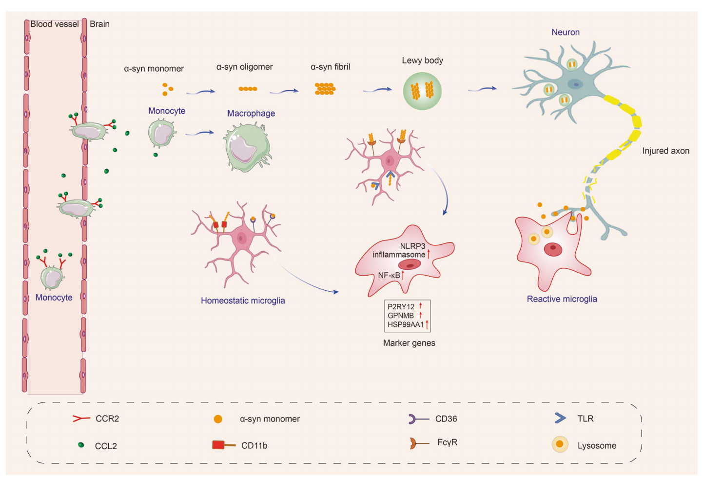
2.1 Neuroinflammation and Microglial Activation: In the substantia nigra of PD patients, a significant accumulation of activated microglia is observed, along with elevated levels of proinflammatory cytokines such as TNF-α and IL-1β. These inflammatory cascades exacerbate neuronal damage. Activated microglia express TREM2, TLRs, and other receptors that sense pathological cues, leading to downstream activation of NF-κB and the NLRP3 inflammasome, perpetuating chronic neuroinflammation.
Kraft et al. (2012) reported that inhibition of TLR4 in an MPTP-induced PD mouse model reduced neuroinflammation and preserved dopaminergic neurons. Houser et al. (2019) further demonstrated that NLRP3 knockout mice exhibited attenuated inflammatory responses and neuronal loss in PD models, highlighting the critical role of inflammasomes in PD pathology.
2.2 α-Synuclein Aggregation:A pathological hallmark of PD is the extracellular accumulation of α-synuclein (α-syn), which can be recognized and phagocytosed by microglia. However, impaired autophagic function compromises this clearance capacity. Excess α-syn can also induce microglial activation and neurotoxicity, creating a vicious cycle. Russo et al. (2019) found that while wild-type microglia efficiently internalize α-syn fibrils, TREM2-deficient or LRRK2-mutant microglia exhibit markedly reduced phagocytic efficiency.
Fig1. α-syn resulting in microglial response in Parkinson’s disease.(Gao, C et al., 2023)
3. Microglial Autophagy and PD
Autophagy is a fundamental cellular process for degrading damaged organelles and aggregated proteins. In microglia, autophagy is regulated via classical pathways involving ATG proteins or noncanonical routes mediated by Rubicon. In PD, the expression of autophagy-related genes such as Atg5 and BECN1 is reduced, impairing microglial autophagy and leading to the accumulation of pathological proteins and heightened inflammation. For instance, DJ-1-deficient microglia exhibit defective autophagy and reduced α-syn clearance, contributing to increased neurotoxicity (Nash et al., 2017).
Heckmann et al. (2019) showed that Rubicon deletion in microglia restored noncanonical autophagy in the 5xFAD model, enhancing Aβ and α-syn clearance. Tu et al. (2021) demonstrated that Atg5 knockout in microglia exacerbated neuroinflammation and dopaminergic neurodegeneration.
Furthermore, the AMPK-ULK1 pathway has emerged as a key regulator of autophagy initiation. Lee et al. (2019) reported that pharmacological activation of AMPK upregulates LC3 expression and improves autophagic flux in microglia, offering a potential therapeutic strategy for PD.
4. Microglial Interactions in PD
4.1 Microglia-Neuron Interactions: Microglia influence neuronal plasticity via synaptic pruning and neurotrophic support. In PD, overactivated microglia release glutamate and reactive oxygen species (ROS), leading to neuronal apoptosis. Fricker et al. (2018) showed that ROS produced via NADPH oxidase damages neuronal DNA under inflammatory conditions.
4.2 Microglia-Astrocyte Interactions: Activated microglia secrete IL-1α, TNF-α, and C1q, inducing astrocytes to adopt a neurotoxic A1 phenotype. Liddelow et al. (2017) reported increased expression of C3 and Serping1 in A1 astrocytes in PD models, indicating their detrimental role.
4.3 Exosome-Mediated Pathology Spread:Microglial exosomes containing α-syn propagate pathology to other brain regions. Guo et al. (2020) demonstrated that labeled microglial exosomes travel between the striatum and cortex, triggering local inflammation and contributing to disease spread.
5. Therapeutic Approaches Targeting Microglia
5.1 Enhancing Autophagy: Agents that activate AMPK, upregulate BECN1, or inhibit Rubicon can restore microglial autophagy and promote protein clearance. For instance, Zhou et al. (2021) developed the AMPK activator C13, which effectively reduced α-syn levels and inflammation in PD models.
5.2 Suppressing Proinflammatory Pathways:Targeting TREM2, TLR/NF-κB, or NLRP3 with small molecules or antibodies mitigates inflammation. MCC950, an NLRP3 inhibitor, suppresses α-syn aggregation and dopaminergic degeneration, lowers IL-1β levels, and improves behavioral deficits (Gordon et al., 2018).
5.3 Phenotypic Reprogramming: Vitamin D promotes microglial polarization toward the M2 phenotype, marked by CD163 and CD206 expression, conferring anti-inflammatory and neuroprotective effects. Cytokines such as IL-4 and IL-10 also induce M2 transition. Chen et al. (2016) demonstrated that IL-10 inhibits M1 polarization in LPS-stimulated microglia.
5.4 Microbiota Modulation: Gut microbiota influence microglial states. Sampson et al. (2016) showed that fecal microbiota transplantation from PD patients to germ-free mice induced motor deficits, underscoring the gut-brain axis’s role in PD.
5.5 TREM2 Gene Intervention Activation: Targeted interventions to enhance TREM2 gene activity in microglia have shown potential to boost Aβ and tau clearance, reducing neuroinflammation and disease progression. TREM2 agonist antibodies or gene delivery systems that increase TREM2 expression improve microglial response to pathological proteins, suggesting that TREM2 activation could be a pivotal mechanism in neurodegenerative disease treatment (Gao, C et al., 2023). Brain Case has introduced an advanced AAV11 serotype vector specifically engineered for high-efficiency and microglia-specific transduction. This vector enables precise genetic manipulation, including targeted gene overexpression or knockdown, and is compatible with the integration of cutting-edge technologies such as optogenetics, chemogenetics, and calcium imaging. It offers a robust and versatile platform for elucidating the functional roles, underlying mechanisms, and pathological contributions of microglia in neurological disorders.
Microglia occupy a central position in the pathogenesis of Parkinson’s disease. Their roles in autophagy, inflammation regulation, intercellular interactions, and pathological propagation offer new insights into PD. Future studies should focus on elucidating microglial heterogeneity, dynamic transcriptional profiles, and spatiotemporal behavior to facilitate precise modulation and targeted therapy. With the advancement of multi-omics technologies and high-resolution imaging tools, microglia-targeted interventions hold promising potential for PD treatment.
Ref:
Masuda T, et al. Spatial and temporal heterogeneity of mouse and human microglia at single-cell resolution. Nature, 2019.
Kraft AD, et al. Toll-like receptor 4-deficient mice are protected from MPTP-induced dopaminergic neurodegeneration. Brain Res, 2012.
Houser MC, et al. NLRP3 Inflammasome Modulation as a Therapeutic Strategy in Parkinson's Disease. Neurotherapeutics, 2019.
Russo I, et al. TREM2 is associated with enhanced microglial phagocytosis of amyloid-beta but not tau aggregates. Alzheimers Res Ther, 2019.
Gao, C., Jiang, J., Tan, Y., & Chen, S. (2023). Microglia in neurodegenerative diseases: mechanism and potential therapeutic targets. Signal Transduction and Targeted Therapy, 8(1), 359.
Nash Y, et al. DJ-1 deficiency impairs autophagy and reduces alpha-synuclein clearance by microglia. J Neuroinflammation, 2017.
Heckmann BL, et al. Noncanonical function of an autophagy pathway suppresses a pathological immune response. Nature, 2019.
Tu Z, et al. Microglial Atg5-deficiency aggravates dopaminergic neurodegeneration in a Parkinson's disease model. Cell Death Dis, 2021.
Lee JH, et al. AMPK mediates autophagy activation to protect neuronal cells from oxidative stress. Biochem Biophys Res Commun, 2019.
Fricker M, et al. Mitochondrial ROS and NADPH oxidase drive microglial phagocytosis of apoptotic neurons. Cell Death Differ, 2018.
Liddelow SA, et al. Neurotoxic reactive astrocytes are induced by activated microglia. Nature, 2017.
Guo M, et al. Microglial exosomes facilitate α-synuclein transmission in Parkinson's disease. Acta Neuropathol, 2020.
Zhou J, et al. C13, a novel AMPK activator, alleviates Parkinson's disease pathology. Neuropharmacology, 2021.
Gordon R, et al. Inflammasome inhibition prevents alpha-synuclein pathology and dopaminergic neurodegeneration in mice. Sci Transl Med, 2018.
Chen Z, et al. Interleukin-10 modulates microglial polarization and reduces neuroinflammation in a LPS-induced model. J Neuroinflammation, 2016.
Sampson TR, et al. Gut microbiota regulate motor deficits and neuroinflammation in a model of Parkinson’s disease. Cell, 2016.
We are very interested in collaborating with your laboratory to expand the applications of our serotype. If you are interested, please feel free to contact us at BD@ebraincase.com