Epilepsy-an ancient neurological disorder-Brain Case ()
Epilepsy, an ancient neurological disorder
Epilepsy (Epilepsy) is one of the common diseases of the nervous system. Patients often present with sudden and short-term abnormalities in movement, sensation, consciousness or spirit. Repeated seizures seriously affect the patient's life and psychology, and even impair cognitive function. Data show that the quality of life of patients with epilepsy is significantly lower than that of the normal population, and lower than that of patients with other chronic diseases, such as hypertension and diabetes. According to epidemiological surveys, the annual prevalence rate of the general population is 5‰~7‰[1], and the prevalence rate of active epilepsy (with seizures within 5 years) is 4.6‰. At present, there are more than 9 million epilepsy patients in my country, of which 5 million to 6 million are patients with active epilepsy, and the number of new patients each year is 650,000 to 700,000. However, its pathogenesis is very complex and has not been fully elucidated so far. Recent studies have shown that epilepsy is closely related to ion channels, neurotransmitters, glial cells, contact transmission and gap junctions.
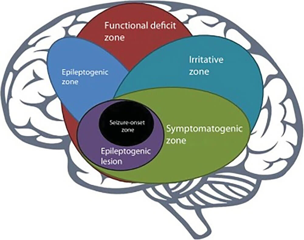
Figure 1. Overlap of cortical areas associated with seizures.
Epilepsy is a chronic encephalopathy characterized by epileptic seizures. Epilepsy syndromes are defined as epilepsy syndromes that have special etiology, specific pathogenesis, special clinical manifestations, and require special treatment. The International League Against Epilepsy defines "epilepsy syndrome" as a group of special manifestations composed of typical clinical and EEG features supported by specific etiology (structural, genetic metabolism, immunity and infection) [2-6]. Manifestations are usually age-dependent and have a specific set of complications.
Currently widely used is the 1985 International League Against Epilepsy classification criteria for epilepsy syndromes [7]. With the advancement of medical technology, especially the development of quantitative EEG and fMRI, and the in-depth understanding of epilepsy neural networks, the International League Against Epilepsy will propose a new classification of epilepsy syndromes in 2022. The new classification still follows the basic framework of the previous classification according to age, and is divided into four categories: onset in neonatal period and infancy, onset in childhood, onset in different ages, and primary hereditary epilepsy syndrome. The classification is refined for clinical application [2-6,8].
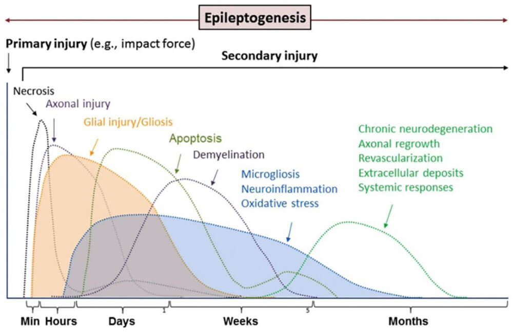
Figure 2: Pathological changes progress and change with the development of epileptogenesis and epilepsy.
Ion channels are the structural basis for the regulation of excitability in excitable tissues in vivo, and are closely related to the occurrence of epilepsy.Some studies have shown that epilepsy is an ion channel disease, which may be due to mutations in the gene encoding ion channel proteins, resulting in changes in ion channel function, resulting in abnormal excitability or inhibitory changes in nervous tissue, and then leading to epileptic seizures[9]. The function of ion channels is to selectively allow corresponding ions to pass through. Currently, it has been found that ion channels related to epilepsy include sodium, potassium, and calcium ion channels. After ions pass through ion channels, they will cause changes in the cell membrane potential, resulting in neuronal excitation or inhibition. Benedictus MR et al. [10] found that ion exchange imbalance can also cause ion channel diseases, that is, epilepsy induced by anions or cations. Therefore, most cases of epilepsy result from abnormal changes in electrolyte distribution and transport, leading to ion channel dysfunction, which in turn causes neuronal excitation and triggers epileptic seizures.
Currently, antiepileptic treatment works by increasing the cerebrospinal fluid γ-aminobutyric acid (GABA) levels in epilepsy patients, thereby altering the corresponding ion channels to generate early inhibitory postsynaptic potentials. Through its excitatory or inhibitory effects, this approach helps control epileptic seizures. Therefore, GABA receptors are involved in epilepsy, and ligand-gated chloride ion channel receptors may also be associated with seizures, although their precise mechanisms of action remain unclear. In summary, epileptic seizures are closely related to neural imbalance, particularly in connection with GABA levels and glutamate receptors[11].
Hanak TJ et al. [12] conducted a study on the immune function of epilepsy patients, showing that the rate of immune dysfunction in epilepsy patients was significantly higher than in other populations. Additionally, the levels of T3, T4, and the T4/T8 cell ratio were lower than those in the normal population, while T8 cell levels were higher (p > 0.05), suggesting a possible link between immune response and epileptic seizures. Inflammatory factors are key regulators of immune and inflammatory responses, indicating that changes in their levels may affect neuronal degeneration, thereby triggering epileptic seizures. Therefore, epileptic seizures are closely associated with immune responses and inflammatory cytokines, although the precise mechanisms of their involvement require further investigation.
Epilepsy has a complex relationship with genetics, and may be associated with a variety of genetic changes. Some patients with epilepsy syndrome may be caused by excessive neuronal excitation caused by mutations in genes encoding ion channel proteins, and gene mutations may include single or multiple gene mutations, chromosomal abnormalities, mitochondrial mutations, etc. However, the specific pathological mechanism remains to be further explored. Although the development of modern molecular biology can provide references for the susceptibility genes of epilepsy-related polygenic genetic diseases, the environmental factors are still difficult to predict.
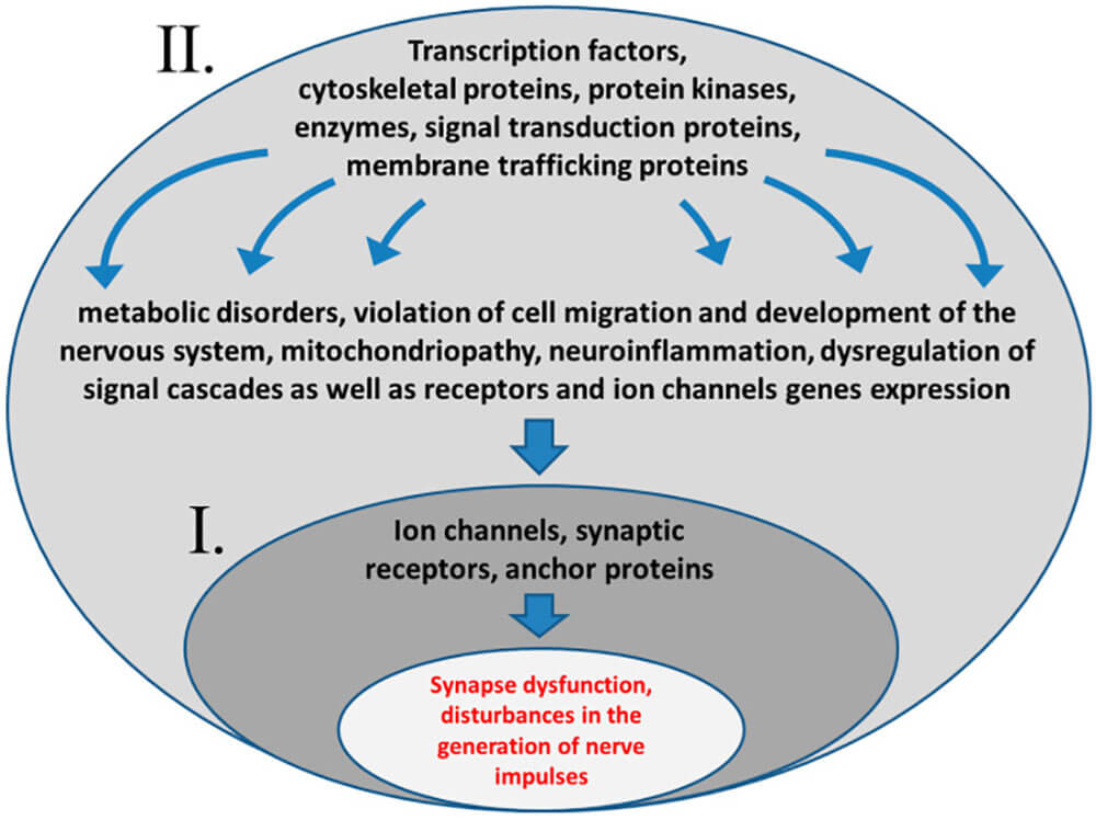
Figure 3: Mutations that lead to the development of epilepsy may affect genes directly involved in the generation and transmission of nerve impulses (I), or affect genes responsible for nervous system development and metabolism (II). In the second case, the violation of the functions of ion channels and specific synaptic proteins occurs indirectly.
Animal models of epilepsy
At present, there are many experimental animal models of epilepsy, which can be classified in many ways according to their seizure conditions, seizure types, and preparation methods, but there is no one model that can perfectly simulate various types of human epilepsy.
Category
Model Name
Introduction
Advantages/Disadvantages
chemistry medicine
Pentylenetetrazol,
PTZ
The most classic chemical drug is to block GABA to transmit pathogenic PTZ, which can be used to build animal models of acute and chronic epilepsy.
kainic acid,KA
Intracerebral injection of KA can be used to study the effect of epileptogenic focus on surrounding and normal brain tissue, while systemic injection of KA. KA is commonly used to study the susceptibility of multiple regions of the brain to epileptogenesis and drug research.
The KA model exhibits similarities with humans in terms of latency period, behavioral symptoms, duration of latency period, and EEG characteristics during both the latency and chronic phases.
Pilocarpine
The ability of pilocarpine (pilocarpine) to induce epilepsy may depend on the activation of M1 receptors, and can cause the increase of glutamate levels in the hippocampus and the activation of NMDA receptors after seizures, thereby maintaining seizures.
The model has recurrent spontaneous seizures, typical hippocampal sclerosis moss fiber germination and other characteristics. Pilocarpine model compared to the above-mentioned drug attacks are more durable, more reliable, and the experimental time shorter and lowest cost.
Penicillin
The penicillin model is more commonly used to mimic drug-resistant absence seizures, which can be exacerbated by PTZ, photostimulation, and GABAergic agonists.
However, when intramuscularly injected into rodents, it does not continuously produce bilateral synchronous spike wave discharges (SWDs) similar to cats, but instead produces multifocal spikes.
However, pretreatment of rats with penicillin can increase the induction of SWDs by other chemicals [17], which is very useful for studying the cellular mechanisms related to spike-wave bursts and the role of cortex on subcortical structures when spike-wave bursts are generated.
Low-dose penicillin-induced focal epilepsy model is suitable for research analysis of the spread of seizure activity in epileptic seizures.
AY-9944
AY-9944 is a compound that inhibits the degradation of 7-dehydrocholesterol into cholesterol. It is the only permanent model of atypical absence. Its induction mechanism is not yet fully understood.
This model has advantages over genetic models in terms of reliability, spontaneity, relapse, and chronic seizures that induce dementia. However, the EEG monitoring required to identify and quantify experimental absence seizures is more demanding.
Hydroxybutyrate,
GHB
The use of biologically active GHB precursor (GBL) produces the exact same EEG and behavioral effects as GHB, but the effects are relatively more sustained and rapid.
The GHB model is a recognized model of absence seizures, and double
Laterally synchronized with cessation of behavior, facial myoclonus, and tremor jerks Twitch related SWDs.
Physical method
Electrical stimulation
The use of biologically active GHB precursor (GBL) produces the exact same EEG and behavioral effects as GHB, but the effects are relatively more sustained and rapid.
This model exhibits a high similarity in both overt manifestations and EEG patterns to psychomotor seizures. It is also considered a screening tool for potential therapeutic drugs for drug-resistant epilepsy. However, it is unrelated to hippocampal sclerosis, and the damage is dissimilar to temporal lobe epilepsy. Spontaneous seizures occur relatively infrequently compared to drug-induced ones.
Low temperature
The low temperature injury model is a model that can induce focal epilepsy without injecting exogenous drugs into the body.
The seizures can last for several days, are repeatable, and have a low mortality rate (<5%). Spontaneous seizures can occur, but the effects of animal age and systemic administration on seizure threshold in this model are not yet clear.
Genetic model
Several neuronal nicotinic acetylcholine receptor genes (CHRNA4, CHRNB2, CHRNA2) and KCNT1 cause autosomal dominant nocturnal frontal lobe epilepsy and LGI1 in rodent models with autosomal dominant epilepsy with auditory features.
Neonatal Convulsion Model
Hypoxia-ischemia,HI
The rapid ischemia-hypoxia model replicated some structural abnormalities and cognitive impairments seen in neonatal seizure. However, there is no obvious brain damage in this model, and differences between strains are minimal.
The HI model typically does not induce seizures in younger (P5) and adult (P60) rats, which may involve other mechanisms.
Febrile seizures,FSs
FS does not involve significant neuronal loss, and seizures in animals are induced by hyperthermia, which differs from the disease process in humans.
Epilepsy Model Evaluation
Symptom Grading of Epilepsy Models - Racines Epilepsy Behavioral Scale
Level 0: normal behavior;
Grade I: Facial muscle spasm manifested as chewing movements, eye blinking, vibrissae, etc., wet dog shaked;
Grade II: neck muscle spasm manifested as a nodding movement;
Grade III: one-sided forelimb clonus;
Grade IV: Standing with bilateral forelimb clonus;
Grade V: On the basis of Grade 4, the body falls backward and loses balance, and the limbs twitch.
This grading later became a unified standard for observing the seizure behavior of the epilepsy model and judging whether the model is successful. Now this standard has been widely adopted by scholars from all over the world.
There are two criteria for judging whether an animal model of epilepsy meets human epilepsy:
The bioelectrical discharge and seizure behavior of the model must be consistent with human epilepsy;
If there is no seizure symptom, EEG should show abnormal discharge of bioelectricity, which is called epileptiform discharge.
In addition to the above-mentioned characteristics, an animal model of epilepsy should be resistant to most antiepileptic drugs, and neuropathologically specific hippocampal damage is related to hippocampal sclerosis in temporal lobe epilepsy.
Outlook
Taken together, these animal models have played a pivotal role in uncovering the underlying mechanisms of epilepsy, determining the efficacy of drugs to treat specific epilepsy types, and preventing the development of epilepsy. However, it is difficult for us to design an animal model that can summarize all the characteristics of epilepsy, and the current models are not perfect, so we should be aware of the limitations of various models. But it needs to be emphasized that animal models should be simple representations of complex systems, mimicking specific aspects of disease, and by no means attempt to replicate all the complexities of human disease. Therefore, whenever an animal model is used, it is critical to define a specific problem and ensure that the chosen model is fit for purpose, making and selecting a model for the purpose.
Literature citation 1. Ure JA, Perassolo M. Update on the pathophysiology of the epilepsies. Journal of the Neurological Sciences. 2000;177(1):1-17. doi:10.1016/S0022-510X(00)00356-7
2. Specchio N, Wirrell EC, Scheffer IE, et al. International League Against Epilepsy classification and definition of epilepsy syndromes with onset in childhood: Position paper by the ILAE Task Force on Nosology and Definitions. Epilepsia. 2022;1398-1442. doi:10.1111/epi.17241
3. Zuberi SM, Wirrell E, Yozawitz E, et al. ILAE classification and definition of epilepsy syndromes with onset in neonates and infants: Position statement by the ILAE Task Force on Nosology and Definitions. Epilepsia. 2022;1349-1397. doi:10.1111/epi.17239
4. Riney K, Bogacz A, Somerville E, et al. International League Against Epilepsy classification and definition of epilepsy syndromes with onset at a variable age: position statement by the ILAE Task Force on Nosology and Definitions. Epilepsia. 2022;1443-1474. doi:10.1111/epi.17240
5. Hirsch E, French J, Scheffer IE, et al. ILAE definition of the Idiopathic Generalized Epilepsy Syndromes: Position statement by the ILAE Task Force on Nosology and Definitions. Epilepsia. 2022;1475-1499. doi:10.1111/epi.17236
6. Wirrell EC, Nabbout R, Scheffer IE, et al. Methodology for classification and definition of epilepsy syndromes with list of syndromes: Report of the ILAE Task Force on Nosology and Definitions. Epilepsia. Published online May 3, 2022. doi:10.1111/epi.17237
7. Chen Y, Li S. Epilepsy, Status Epilepticus, and Refractory Status Epilepticus. Refractory Status Epilepticus. 2017;1-42:1-41. doi:10.1007/978-981-10-5125-8_1
8. Wirrell E, Tinuper P, Perucca E, Moshé SL. Introduction to the epilepsy syndrome papers. Epilepsia. 2022;1330-1332. doi:10.1111/epi.17262
9. Van Bogaert P. KCTD7-related progressive myoclonus epilepsy. Epileptic Disorders. 2016;18(S2):115-119. doi:10.1684/epd.2016.0856
10. Benedictus MR, Prins ND, Goos JDC, Scheltens P, Barkhof F, van der Flier WM. Microbleeds, Mortality, and Stroke in Alzheimer Disease. JAMA Neurology. 2015;72(5):539. doi:10.1001/jamaneurol.2015.14
11. Peljto A, Barker‐Cummings C, Vasoli V, et al. Familial risk of epilepsy: a population-based study. Brain : a journal of neurology. 2014;795-805. Accessed December 20, 2022. https://www.semanticscholar.org/paper/Familial-risk-of-epilepsy%3A-a-population-based-Peljto-Barker%E2%80%90Cummings/fecc1c896ceb0e0a8a7606deec6afb698a540a1b
12. Hanak TJ, Libbey JE, Doty DJ, Sim JT, DePaula-Silva AB, Fujinami RS. Positive modulation of mGluR5 attenuates seizures and reduces TNF-α+ macrophages and microglia in the brain in a murine model of virus-induced temporal lobe epilepsy. Experimental neurology. 2019;311:194-204. doi:10.1016/j.expneurol.2018.10.006
One-step Service
In the field of drug efficacy and pharmacological evaluation related to neurological diseases, Brrain Case can provide you with a one-stop platform for evaluating the behavior of animals from the gene molecular level to the cell tissue level, to the neural circuit, and finally to the animal as a whole.
At the gene molecular level, through gene editing, gene interference, in situ hybridization, immunohistochemistry and other technical means, verify the influence of genes or proteins on cell physiological metabolic signal transduction, gene expression regulation, etc. Experimentally verify the function of a molecule.
At the neural circuit level, we can analyze the structure and function of neural circuits formed between different brain regions and different types of neurons by means of circuit tracing, optogenetics, chemical genetics, and electrophysiology. Such research is one of the key development directions of neuroscience at present. Understand, manipulate and analyze the dramatic phenotypic differences brought about by changes in neuronal connectivity.
At the same time, Brain Case also provides a high-precision animal behavior testing platform after neurological disease modeling, including but not limited to: cognitive function testing, motor function testing, multi-channel in vivo electrophysiological recordings of awake animals, respiratory recordings, auditory, Pain, anxiety, depression, behavioral tests related to olfactory function, etc.
MRI/fMRI/PET-CT small animal living imaging, cardiac ultrasound, X-ray film, tissue fluorescence imaging (whole brain slide scanning, tissue immunofluorescence imaging), calcium imaging, and confocal two-photon imaging can also be provided.
If you are interested in building animal models of epilepsy and evaluating drug efficacy after modeling or administration, please feel free to contact us at BD@ebraincase.com