An important method for detecting neuronal activity: calcium signals recording
It is a method that uses calcium ion indicators/probes to detect changes in calcium signals in tissues. Calcium ion imaging technology is mainly used in nervous system research, where changes in calcium ions indicate neuronal activity. This technology is widely used in the fields of neurobiology, cell biology, physiology, developmental biology and pharmacology. Among them, neurobiology mainly studies the signal emission caused by changes in ion concentration of nerve cells, physiology mainly studies muscle movement-calcium signals in cardiomyocytes, cell biology mainly studies signal transduction and ion channels, and developmental biology mainly studies The egg fertilization mechanism is mainly used in pharmacology to screen drugs and investigate pharmacodynamics.
Principle of calcium signals recording technology
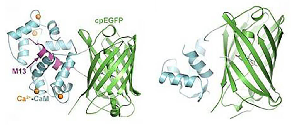
Calcium signals recording technology belongs to a category of optogenetic technology. It actually detects the concentration changes of Ca2+ in cells or tissues and converts the changes in calcium concentration into fluorescence signals, thereby converting cell electrical activity into recordable light signals. The detection method is completed by using a calcium ion indicator that can sense the concentration of Ca2+. Calcium ion indicators are divided into two types, one is chemical calcium ion indicators (chemical calcium indicators), and the other is now commonly used genetically encoded calcium ion indicators (genetically-encoded calcium indicators, GECIs), such as GCaMP . GCaMP is composed of green fluorescent protein (GFP), calmodulin (CaM) and a peptide sequence M13 of myosin light chain kinase. When Ca2+ binds to CaM, CaM undergoes a conformational change, and its light chain region can bind to M13. Under illumination of a specific wavelength, the protonation of the GFP chromophore is enhanced and the absorbance increases, causing GFP to emit strong fluorescence. As shown in Figure 1.
Figure 1. Schematic diagram of the structure and function of GCaMP (from Wikipedia)
Three commonly used methods for calcium signals recording in vivo
1. Calcium ion fiber optic recording (Fiber Photometry)
Calcium ion fiber optic recording is based on fluorescence imaging of calcium ion concentration changes, which can examine and record cell activity changes in real time. The emergence of this technology is of milestone significance for the detection of circuit-level neural signals encoding social behaviors, learning, memory, and fear behaviors, as well as pathological conditions.
Advantages:
Enables cell type-specific activity detection;
Strong resistance to interference;
Easily achieves long-term activity monitoring.
Limitations:
It cannot be used to record the activity of individual neurons.
Fiber optic recording experimental steps :
① Inject a certain amount of GECIs(such as GCaMPs) virus into specific brain areas of experimental animals.
② Implant a fiber-optic ceramic ferrule with a diameter of 200-400 μm at the same location where the GCaMP virus was injected to transmit excitation light and collect emission light.
③ The optical fiber recording system can be divided into a fluorescence excitation system and a fluorescence collection system in principle. The excitation light reaches specific brain areas after passing through the optical fiber jumper and the ceramic ferrule on the animal's head, thereby stimulating the green fluorescence signal. The intensity of the excited fluorescence signal can be collected by the end of the optical fiber implanted in the animal's brain. The collected fluorescence is converted into electrical signals by the sensor, and then transmitted to the recording system through the data acquisition card to achieve real-time observation of a group of brain areas under study. Purpose of neuronal calcium fluorescence activity.
④ Data analysis. Fluorescence signals are usually divided into different segments based on behavior, and aligned neatly with time zero points based on specific behavioral events. We use the difference between the current fluorescence intensity F and the baseline fluorescence intensity F0 and divide it by the value of the baseline fluorescence intensity F0, that is, the value of ΔF/F0 = (F−F0)/F0 to characterize the fluorescence intensity change around the event. The results of analysis are commonly represented by two types of graphs, namely heatmap and peri-event plot.
2. Two-photon in vivo calcium imaging
Using two-photon imaging technology, when in vivo stimulation is given, the light changes of nerve cells labeled by GFP are recorded to determine nerve cell activity. This method is suitable for selecting the surface layer of the brain as the study area.
Advantage:
1) In a two-photon microscope, the wavelength of the excitation light is around 700-1000nm, which is close to the infrared spectrum. This is the first advantage of in vivo two-photon imaging. Longer wavelength light scatters less in tissue and penetrates deeper.
2) Has good resolution, allowing visualization not only of single neurons but also of calcium activity in axonal terminals of subcellular structures.
Shortcomings:
1) Photobleaching that may occur. The excitation light intensity required to distinguish signal from noise usually needs to be high enough to cause some degree of photobleaching, and photobleaching overtime can have a negative impact on image quality.
2) The imaging depth is restricted by many factors. Microendoscopes (Gradient Refractive Index (GRIN) lenses) can be implanted to visualize deeper brain regions, but due to the size of the objective lens, large portions of the cortex or other brain structures often need to be completely removed to allow for lens implantation Leave space as this may seriously affect the animal's behavior.
3) The animal’s head needs to be fixed. It is believed that the head-fixed animals were under physical restraint and emotional stress during the experiment, so it was impossible to prove that the neuronal responses to the outside world in this environment were equivalent to those under free exploration. What's more, many social behaviors, such as parent-child care, mating, and fighting, cannot be studied using head-fixed experiments.
Two-photon in vivo calcium imaging experimental steps:
①In the first surgery, the dura mater is exposed and micro-injection of AAV (50-100 nL, titer approximately 1012) is performed;
②The animals are released back into the cage for recovery and given antibiotics for one week;
③A second surgery will be performed 45-60 days later to implant head posts in the forehead and back of the head, and connect these head posts with a T-shaped steel frame;
④Open the dura mater again to expose the cortex, push the window unit composed of glass cover, silicone glue, titanium ring and GORE membrane to the cortical surface, insert the GORE membrane under the dura mater, and bond the titanium ring and skull to form an imaging cavity;
⑤Perform behavioral tasks or stimuli, and perform two-photon imaging using a two-photon microscope approximately 10 days after recovery from the secondary surgery.
3. Single-photon micromicroscopy for intravital calcium imaging
Advantage
1) Based on single-photon in vivo calcium imaging using micromicroscopes, researchers can record calcium signals in freely moving animals.
2) Through the connection with the gradient refractive index (GRIN) lens, the observation of the activity of group neurons over a long period of time can be completed.
3) The device is small in size and can be fixed on the animal's head without seriously hindering its movement. The development of this technology has made microscopic microscopy a very useful tool for studying development, neuroplasticity and neurodegenerative diseases.
Disadvantages
1) Compared with two-photon imaging, single-photon imaging has higher background fluorescence and is more prone to light scattering.
2) The application is limited in certain behavioral experiments, such as water maze and forced swimming experiments.
3) Since the diameter of the GRIN lens implant ranges from 0.5mm to 1mm, it will inevitably cause a certain range of tissue damage, which may affect the key circuits of the study.
Commonly used genetically encoded calcium indicators (GECIs), see the table below:
Category
Name
Application
References
GCaMP6 series
GCaMP6s
Highly sensitive, suitable for indication of low frequency signals
PMID: 23868258
GCaMp6m
Moderate binding activity, widest range of application
GCaMp6f
Fast kinetic curve, fastest dissociation, suitable for indication of high-frequency signals
jGCaMP7 series
jGCaMP7s
Highly sensitive, the sensitivity can be more than 5 times that of GCaMP6s
PMID: 31209382
jGCaMP7f
Fast kinetics, its reaction speed is 3 times that of GCaMP6s and 5 times that of GCaMp6f. Suitable for stronger detection of single action potential responses or group activity experiments.
jGCaMP7b
Brighter baseline fluorescence, 3 times more sensitive than GCaMP6s. Suitable for detecting neuronal processes or nerve fibers.
jGCaMP7c
The background fluorescence is low, the contrast is high, the signal is clear, and the sensitivity is 2.7 times that of GCaMP6s. Suitable for large-scale imaging.
jGCaMP8 series
jGCaMP8f
Fast kinetics, ~4× faster rise time and ~2.5× faster decay time compared to jGCaMP7f
Blue, green, orange, red. Combined with specific neuron-specific expression methods, it is possible to simultaneously monitor the activities of three different neuron types in specific behaviors in a free-moving state; combined with two-photon microscopy, microstructural and functional imaging is performed to achieve presynaptic and synaptic Simultaneous two-color imaging of posterior structures.
PMID: 31080068
XCaMP-G
XCaMP-O
XCaMP-R
Brain Case offers experimental services including optogenetic and chemogenetic manipulation, fiber photometry for calcium and neurotransmitter signal recording, EEG, EMG, and patch-clamp electrophysiology. These services are well-suited for studies of neural activity and function, as well as for evaluating neuropharmacological and neurotoxicological effects.
If you are interested in the details of the experiment or possible problems and causes during the experiment, please contact: BD@ebraincase.com
Service Type :
Select the service you'd like to purchase.
Order Information(Premade-AAVs)
Please provide us some information about the service you'd like to order.
Order Information(Custom AAV/Lentivirus)
Please provide us some information about the service you'd like to order.
Order Information(Others)
Please provide us some information about the service you'd like to order.