RCAS virus is a member of the retrovirus family derived from the SR-A strain Rous sarcoma virus (RSV), a replicating avian sarcoma-leukemia virus (ASLV) with splicing receptors long terminal repeat (LTR). The src oncogene of RSV was deleted and a polyclonal site was inserted that could stably accommodate up to 2.5kb of inserted fragments (Figure 1). The expression of the inserted gene may be driven by the viral long terminal repeat (LTR) or by an appropriate internal promoter.
Fig.1 Constructs of the RSV and RCAS vector
Principles of the RCAS-TVA system
The RCAS-TVA gene delivery system is based on the RCAS virus specifically recognizing the avian sarcoma leukemia virus subgroup A(ASLV-A) receptor (TVA) to enter and infect cells. TVA is a member of the low-density lipoprotein receptor family, encoded by the tv-a gene. The mRNA transcribed by the tv-a gene is selectively spliced to produce at least two proteins, a transmembrane protein and a GPI-anchored isoform, both recognized by ASLV-A. Mammalian cells lack the gene that codes for TVA and are generally resistant to ASLV-A or RCAS virus infection; However, targeting TVA transgenes to specific cell types or mouse tissues can make these cells vulnerable to ASLV-A based RCAS virus infection. RCAS viruses retain all the viral genes needed for replication, and when the RCAS vector enters the cell, it can directly encode the infectious particles and assemble them into the desired proteins, so no helper cells are needed. High titer RCAS virus can be obtained by transfecting RCAS vector directly into avian cells, such as DF1 cells. The generation process based on RCAS-TVA model is shown in Figure 2 [1]. The expression of specific proto-oncogenes by RCAS can induce specific types of cells or mouse tissues to become cancerous and form tumors.
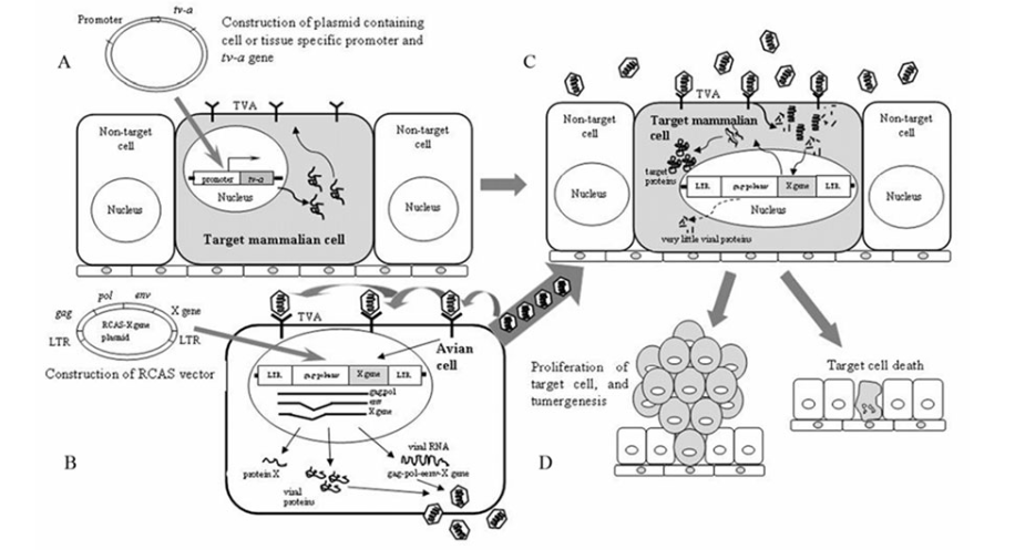
Fig.2 Schematic drawing ofthe RCAS-TVA technique in mammalian system
Advantages and disadvantages of the RCAS-TVA system
1. The RCAS-TVA system is a low-cost, simple and efficient method that can simultaneously study the biological functions of multiple genes using a single TVA mouse strain, such as the influence of multiple genes on tumorgenesis; There is no need to generate a mouse strain from each gene of interest through traditional transgene/knock-out/knock-in methods or multiple rounds of hybridization. For example, in mouse strains with elastase promoter mediated TVA expression, delivery of RCAS-c-myc or RCAS-PyMT can be used to induce three different pancreatic tumor types, also reflecting the three main cell lines in the pancreas [2].
2. The RCAS-TVA system can target specific cell types through TVA and regulate gene expression in time and space through the timed delivery of RCAS virus. This is important for using mouse models to simulate human cancer. Human cancers almost universally occur in single-cell populations that have received multiple gene strikes. These cell clones are physically separated from each other and surrounded by normal cells. Therefore, it is important to model this situation to understand how cancer cells communicate with the surrounding normal cells and micro-environment, and how cancer cell clones can endanger the normal cells and/or micro-environment around them for their own growth advantage. Most human cancers are diseases of aging, so it is ideally important to simulate cancer in adult mice in a controlled manner. For example, a series of studies on breast tumors provide a good example of using the RCAS-TVA system to regulate gene expression in time and space. MMTV-TVA transgenic mice express TVA in the mammary duct epithelium, and inject the RCAS virus expressing polyoma virus T antigen (PyMT) into the mammary gland through the papillary duct to induce breast tumors [3]. For example, regarding the effect of pregnancy on human breast cancer, MMTV-tva mice were transfected by intraductal injection of RCAS-caErbB2 virus 4-7 days before mating, and this breast cancer was diagnosed during human pregnancy or within 5 years after delivery [4].
3. Despite the obvious advantages of using the RCAS-TVA gene delivery system to simulate human cancer in mice, there are certain limitations. First of all, the capacity of RCAS virus is limited, allowing the size of the inserted fragment to be about 3 kb. If the fragment is too large, the inserted fragment may be lost, truncated, or the virus production may be reduced. Another limitation is that RCAS viruses can only integrate viral cDNA into the host genome when the cell is in a dividing state, which greatly limits the number of cells that can be infected in adult tissue, and there may be artificial effects by selectively infecting dividing cells that may not fully represent the properties of other resting cells. An ALSV-A pseudolentivirus has also been developed to infect hard-to-target cell types and tissues using the lentiviral system (Lewis et al. 2001).
Sum up
The RCAS-TVA system provides a simple and rapid way to study gene function in mouse models of human cancer. These models provide important insights into the cells of origin of specific tumors and the role of initial genetic lesions in determining tumor types. The RCAS-TVA model can be combined with existing transgenic, knock-out or knock-in models, providing opportunities to extend the utility of these models.
If you're interested in the modification and application of RCAS, please click on the small window in the bottom right corner or send an email to BD@ebraincase.com to contact us.
Reference 1)Ahronian L G, Lewis B C. Using the RCAS-TVA system to model human cancer in mice[J]. Cold Spring Harbor Protocols, 2014, 2014(11): pdb. top069831. 2)Lewis B C, Klimstra D S, Varmus H E. The c-myc and PyMT oncogenes induce different tumor types in a somatic mouse model for pancreatic cancer[J]. Genes & development, 2003, 17(24): 3127-3138. 3)Holloway K R, Sinha V C, Toneff M J, et al. Krt6a-positive mammary epithelial progenitors are not at increased vulnerability to tumorigenesis initiated by ErbB2[J]. PLoS One, 2015, 10(1): e0117239. 4)Haricharan S, Hein S M, Dong J, et al. Contribution of an alveolar cell of origin to the high-grade malignant phenotype of pregnancy-associated breast cancer[J]. Oncogene, 2014, 33(50): 5729-5739. 5)Lewis B C, Chinnasamy N, Morgan R A, et al. Development of an avian leukosis-sarcoma virus subgroup A pseudotyped lentiviral vector[J]. Journal of virology, 2001, 75(19): 9339-9344.