- E-mail:BD@ebraincase.com
- Tel:+8618971215294
The brain's neural network is a complex structure composed of a large number of neurons with different shapes and functions connected through synapses. It is the structural basis for the brain's cognitive, emotional, memory and imagination activities. It is suggested that the structure of the brain's neural network is the prerequisite for understanding the mechanism of the brain's information processing. The traditional tracer method has promoted people's understanding of the structure of the brain's neural network, but it is difficult to be used to study multiple brain regions and multiple types of neurons through synapses. Connections form complex neural networks, which are the circuits that the brain relies on to process specific information.
Analyzing the neural circuit connections between different brain regions and different types of neurons is one of the important tasks of neuroscience research. Drawing a map of the brain connectome is crucial to understanding how the brain works. Virus tools are currently the most effective, The most widely used neural circuit tracing tool. Neural circuits are the basic units of neural function and are the bridge connecting large scale (structure/function) and small scale (molecules/signaling pathways). Effective mapping of neural circuits requires simultaneous transmission of multi-level or single-level processes by retrograde and anterograde tracer viruses. touch. Currently, viral vector tools that span multiple levels in the forward direction include herpes simplex virus (HSV) and vesicular virus (VSV).

Figure 1. Schematic diagram of neuron orthodromic labeling across multiple levels.
H129 is a clinical isolate of HSV-1 virus that has strict anterograde transsynaptic properties in both the peripheral nervous system and the central nervous system. After the virus infects nerve cells, it replicates and expresses the target gene in the cell. The progeny virus is transported to the presynapse through the axon, crosses the synapse and enters the downstream neuron, starting a new round of replication and cross-synaptic propagation. The modified virus, after being loaded with fluorescent proteins, can achieve efficient and stable anterograde trans-synaptic labeling of neuron networks, which is suitable for anterograde tracing studies of output neural circuit.
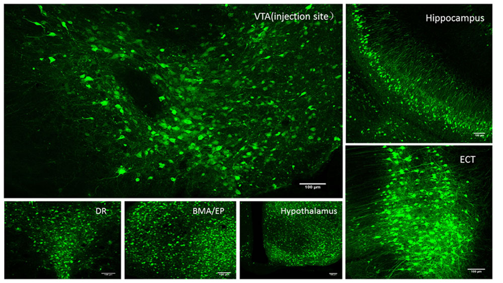
H129-EGFP was injected into the ventral tegmental area (VTA) of mice, and H129-EGFP was observed to travel anterogradely through multiple levels to the hippocampus, dorsal raphe nucleus (DR), dorsomedial anterior segment of the amygdala (BMA), peduncular core (EP), hypothalamus (Hypothalamus), and extranasal cortex (Ect).

Figure 2. Using H129-EGFP to label the output neural circuit of the ventral tegmental area (VTA) of mice
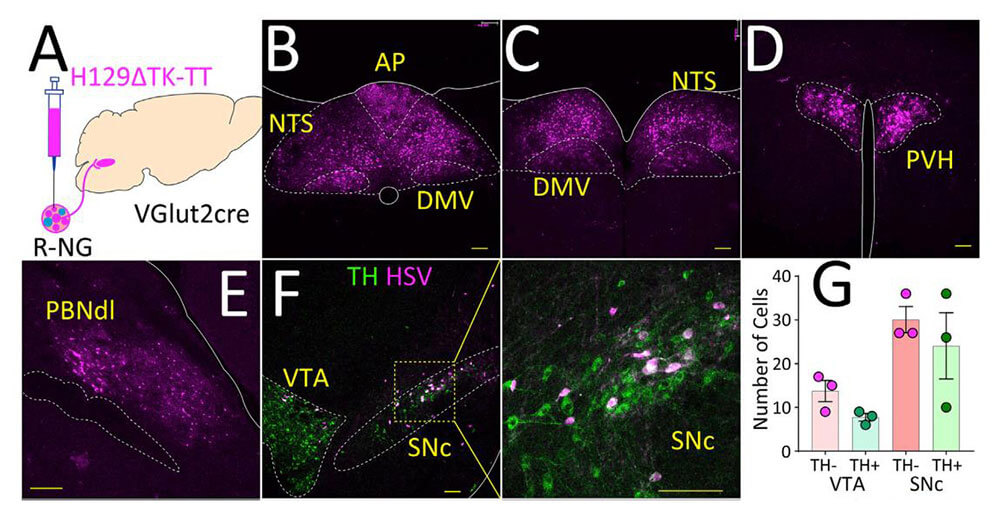
Example 1: The nodose ganglion (NG) anterograde virus tracing across multiple levels

Figure 3. Labeling of central vagal pathways (HanW, et al., Cell, 2018)
VSV virus has specific anterograde transsynaptic properties. When the virus is positioned and injected into the experimental area, the virus infects the nerve cells and replicates in the nerve cells and expresses the fluorescent protein gene it carries. After the progeny virus is produced, it is transported to the presynapse through the axon, crosses the synapse, enters the downstream neuron, and begins A new round of replication, packaging and trans-synaptic propagation processes. The VSV tool virus for anterograde transsynaptic tracing is widely used in neural circuit tracing studies in rodents models. It can also infect a variety of animal models, including fish, birds, and non-human primates. It is characterized by fast replication and trans-synaptic speed, ultra-high expression of foreign genes, and the ability to obtain fine morphology of neurons.
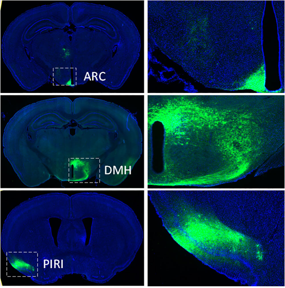
For example, when VSV-EGFP was injected into the Spleen area of C57 mice, fluorescent signals were detected in the ARC, DMH, and PIRI areas of the brain center, indicating that VSV-EGFP traveled anterogradely across multiple levels to these three nuclei.

Figure 4. The output neural circuit of mouse Spleen area injected with VSV-EGFP
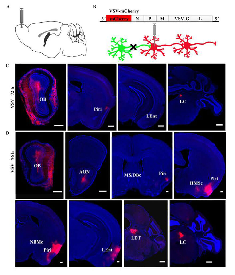
Example 1: OB antrograde muti-synaptic virus tracing

Figure 5. The output neural circuit of the olfactory bulb (OB) to the basal forebrain (BF) (ZhengY, et al., Front Neural Circuitsl, 2018)
| Product No. | Product Name | Fluorescent Protein Carried | Function |
| BC-HSV-HBEGFP | H129-hUbC-HBEGFP | HBEGFP | Enhanced green, antrograde muti-synaptic tracing |
| BC-HSV-EGFP | H129-hUbC-EGFP | EGFP | Green, antrograde muti-synaptic tracing |
| BC-HSV-tdTomato | H129-hUbC-tdTomato | tdTomato | Red,antrograde muti-synaptic tracing |
| BC-VSV-Vs03 | VSV-EYFP(36399) | EGFP | Green,antrograde muti-synaptic tracing |
| BC-VSV-Vs13 | VSV-mCherry(36399mCh) | mCherry | Red,antrograde muti-synaptic tracing |
| Labeling Type | Synaptic Specificity | Common Tools |
|---|---|---|
| Retrograde Tracing | Non-synaptic | AAV2/Re, AAV2/11, RV-ΔG-N2cG, CTB |
| Retrograde Monosynaptic Tracing | Monosynaptic | RV-EnvA-ΔG-XFP |
| Retrograde Mutisynaptic Tracing | Mutisynaptic | PRV-XFP, PRV-△TK-DIO-XFP (Cre-dependent) |
| Anterograde Tracing | Non-synaptic | AAV2/2, AAV2/8, AAV2/9 |
| Anterograde Monosynaptic Tracing | Monosynaptic | AAV2/1, AAV2/9-mWGA, Hs06, H361 |
| Anterograde Mutisynaptic Tracing | Mutisynaptic | HSV, VSV |
| Deliverables (for all services above): Full project report and raw imaging data | ||