- E-mail:BD@ebraincase.com
- Tel:+8618971215294
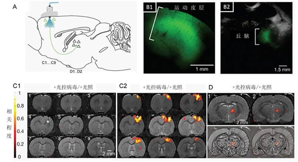
Functional magnetic resonance imaging (fMRI) technology has made great contributions to the development of neurobiology and still has strong potential. The newly developed optogenetics technology (Optogenetics) and the emergence of a new method combined with fMRI - light-activated magnetic resonance functional brain imaging (Optogenetic fMRI, ofMRI, opto-fMRI), provide a more effective way for fMRI technology to promote neurobiology. development provides opportunities. This technology can selectively control the activity of specific types of nerve cells in the brain, realize the activity of one or some specific nodes in a specific brain network, and obtain the response characteristics of the relevant neural network, thereby analyzing the structure and function of the relevant network. , as well as the role and interrelationship of related brain areas in specific brain functions; at the same time, high spatial and temporal precision perturbations and functional hemodynamic responses can be integrated to analyze neural activity, brain energy metabolism and their dependence on blood oxygen levels (Blood Oxygen Level Dependent, BOLD) characteristic relationship between signals.

Figure 1. A. Schematic diagram of virus-infected area and light-stimulated area. B1. Distribution of ChR2-EYFP in the infected area (motor cortex) in situ. B2. Distribution of ChR2-EYFP in the motor cortex projection area, thalamus. C1. The control group without virus infection did not produce positive BOLD signal under light stimulation. C2. Light activates the activity of projection neurons in the motor cortex, and considerable positive BOLD signals are detected in the in situ area (*, photostimulation site). D. Neural activity in the motor cortex can induce BOLD signals in the thalamus. Each layer in C and D is marked in A respectively.Whole-brain BOLD response evoked by light activation of an in situ region infected with a light-controlled virus in the rat cortex (modified from Nature 465, 788-792, May, 2010.
But in fact, the source of the BOLD signal is related to a series of changes in cerebral blood flow, cerebral blood volume, cerebral oxygen consumption metabolic rate, blood vessel density and size, magnetic field strength, etc. On the other hand, the relationship between neural activity and cerebral blood flow, cerebral blood volume and blood oxygen saturation is also quite complex, making the relationship between neural activity and BOLD signal even more complicated. Local hemodynamic changes caused by neural activity are likely to be the combined result of multiple factors. In short, the correlation between neural activity and BOLD is beyond doubt, but the specific mechanism of action between the two still needs to be studied urgently.