- E-mail:BD@ebraincase.com
- Tel:+8618971215294
The brain's neural network is a complex structure composed of a large number of neurons with different shapes and characteristics connected through synapses. It is the structural basis for the brain's cognitive, emotional, memory, imagination and other activities. Mapping neuronal projections can track the flow of information between different brain areas.
Anterograde tracers are mainly taken up by neuronal somata and dendrites and then travel to the axon, thereby marking the area where the neuron projects. Classic anterograde tracers mainly include the following types:
Antrograde tracing is often used with in situ hybridization, immunostaining, or Nissl staining.
A limitation of classical anterograde viral vectors is that all cells at the injection site take up the tracer, so the projection pattern revealed by this method is an overall reflection of the projections from different types of neurons. As a neuron tracking reagent, viruses can be more rigorous in direction, can be combined with Cre-Lox technology to achieve cell type-specific labeling, and can also carry genetic tools (such as optogenetic tools, calcium-sensitive dyes, and gene editing tools, RNA interference tools, etc.) can play an important role in the study of neural circuit functions.
Some commonly used antrograde viral vectors that do not cross synapses include adeno-associated viruses of serotypes 2, 8, and 9, retroviruses, and lentiviruses packaged with VSV-G.
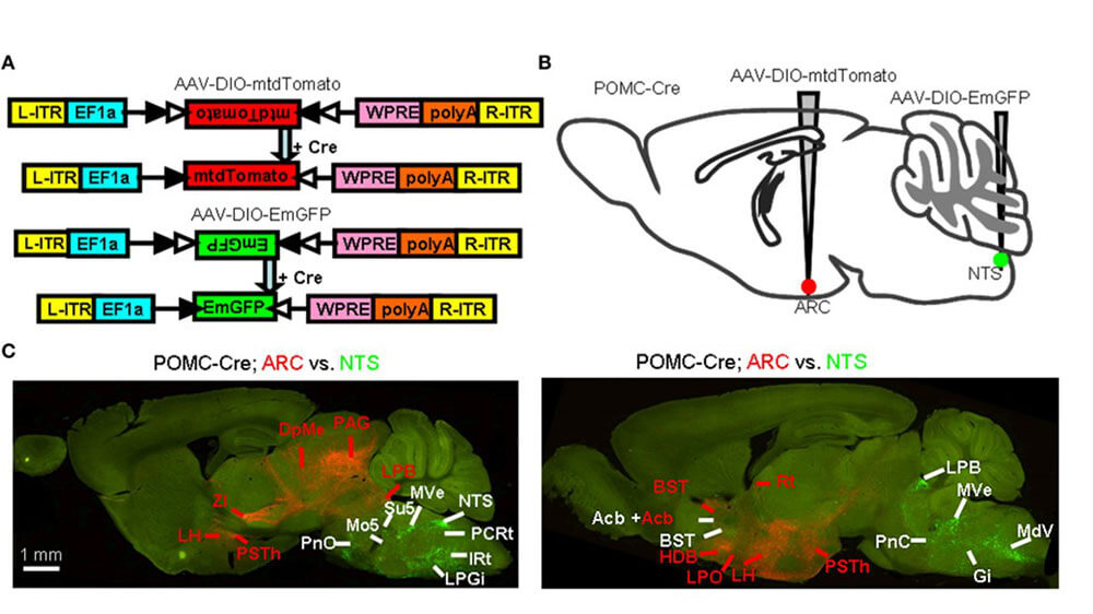
Example 1: Identification of brain-wide efferents from POMC neurons

Figure 1. Identification of brain-wide efferents from POMC neurons (Minmin Luo, et al. Frontiers in Neuroanatomy. 2015)
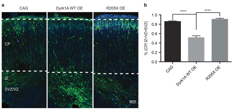
Example 2: The role of Dyrk1a (dual-specificity tyrosine-(Y)-phosphorylation-regulated kinase 1A) in brain development

Figure 2. Dyrk1a(WT) overexpression can delay the migration of neurons in the developing mouse embryonic cortex (ZLQiu, et al. Molecular Psychiatry.2018)
Retrograde tracers are usually taken up by axon terminals and then transported back to the cell body. Classic retrograde tracers include:
Determining whether a tracer is being transported in the forward or reverse direction depends primarily on observation. Retrograde tracers usually bind to receptors that are selectively enriched in axonal terminals, are absorbed through endocytosis at the axonal terminals, and reach the cell body through the endogenous retrograde axonal transport system.
Like the classic anterograde tracers, the above-mentioned traditional retrograde tracers are also directionally non-specific and cannot track specific types of neurons in the same target area.
Some commonly used retrograde viral vectors that do not cross synapses include
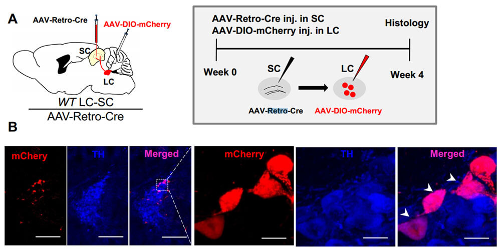
Example 1: Retrograde viral tracing of LC-SC projections

Figure 3. Retrograde viral tracing of LC-SC projections (LipingWang, et al.Current Biology.2018)
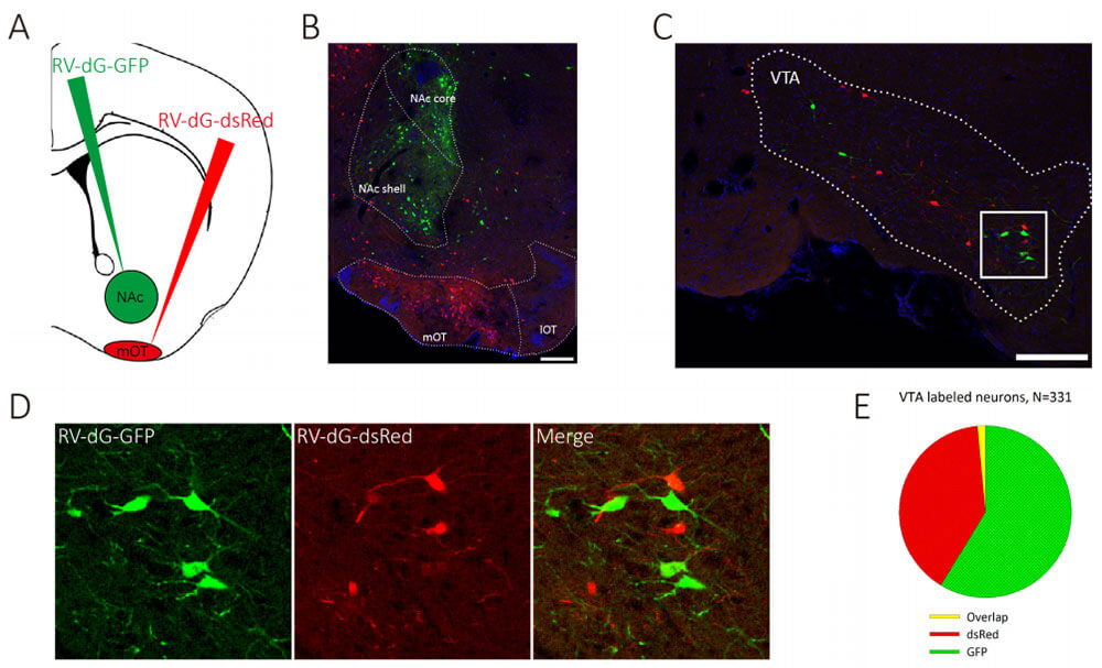
Example 2: Separation of VTAergic neurons innervating NAC and mOT

Figure 4. Isolation of TA-ergic neurons innervating NAC and mOT (Zhijian Zhang, et al.eLife.2017)
| Labeling Type | Synaptic Specificity | Common Tools |
|---|---|---|
| Retrograde Tracing | Non-synaptic | AAV2/Re, AAV2/11, RV-ΔG-N2cG, CTB |
| Retrograde Monosynaptic Tracing | Monosynaptic | RV-EnvA-ΔG-XFP |
| Retrograde Mutisynaptic Tracing | Mutisynaptic | PRV-XFP, PRV-△TK-DIO-XFP (Cre-dependent) |
| Anterograde Tracing | Non-synaptic | AAV2/2, AAV2/8, AAV2/9 |
| Anterograde Monosynaptic Tracing | Monosynaptic | AAV2/1, AAV2/9-mWGA, Hs06, H361 |
| Anterograde Mutisynaptic Tracing | Mutisynaptic | HSV, VSV |
| Deliverables (for all services above): Full project report and raw imaging data | ||