Nature Breakthrough: AAV-ROOT Serotype Enables Precise Tracing of Fat-Sensing Neurons, Challenging Traditional Metabolic Regulation Concepts
Release time:2025-03-12 10:58:28
There exists a complex communication mechanism between adipose tissue and the central nervous system to maintain whole-body energy homeostasis. Traditionally, adipose tissue was thought to convey metabolic state information to the brain through secreted circulating hormones, while the brain regulated adipocyte function via sympathetic nervous outputs. Sensory neurons in the dorsal root ganglia (DRG) have also been shown to innervate adipose tissue, but their physiological significance has remained underexplored due to the lack of specific genetic tools.
《Nature》
On August 31, 2022, the research team led by Li Ye at The Scripps Research Institute published a study in Nature titled "The role of somatosensory innervation of adipose tissues." This study revealed the role of DRG sensory innervation in regulating adipose tissue function, particularly through a novel AAV serotype—ROOT (Retrograde vector Optimized for Organ Tracing)—which enables specific manipulation of adipose tissue innervation.
1. Direct Visualization and Characterization of DRG Projections to Adipose Tissue
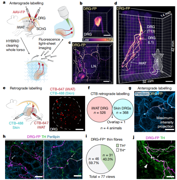
To explore DRG projections within adipose tissue, researchers injected an AAV expressing a fluorescent protein (AAV-FG) into the thoracolumbar DRGs. Utilizing HYBRiD tissue clearing technology and light-sheet microscopy, they directly observed the complete axonal projections of DRG neurons to adipose tissue. The results revealed a large number of FG-positive fibers in the inguinal white adipose tissue (iWAT) (Figure 1a-d). Since iWAT is adjacent to the skin, the researchers next examined whether the DRGs innervating iWAT and the skin were the same. Using dual-color cholera toxin subunit B (CTB) retrograde tracing, they found that DRGs innervating iWAT and those innervating the skin were completely distinct, with no co-labeling observed (Figure 1e-f). These findings further confirmed the direct innervation of adipose tissue by DRG neurons (DRG→iWAT). Furthermore, immunofluorescence and co-localization analysis identified two distinct populations of FG-positive fibers (DRG-FP⁺): Large fiber bundles traveling along blood vessels together with tyrosine hydroxylase (TH)-positive sympathetic fibers (Figure 1j). Sensory nerve terminals closely adjacent to adipocytes, with 40% of them being TH-positive (Figure 1g-h). These findings challenge the conventional view that TH is a specific marker for sympathetic innervation in adipose tissue. The study suggests that previous TH-based research may have been influenced by sensory innervation, at least to some extent.
Figure 1: DRG Innervation of Adipose Tissue
Selective Targeting of Fat-Sensing Neural Innervation
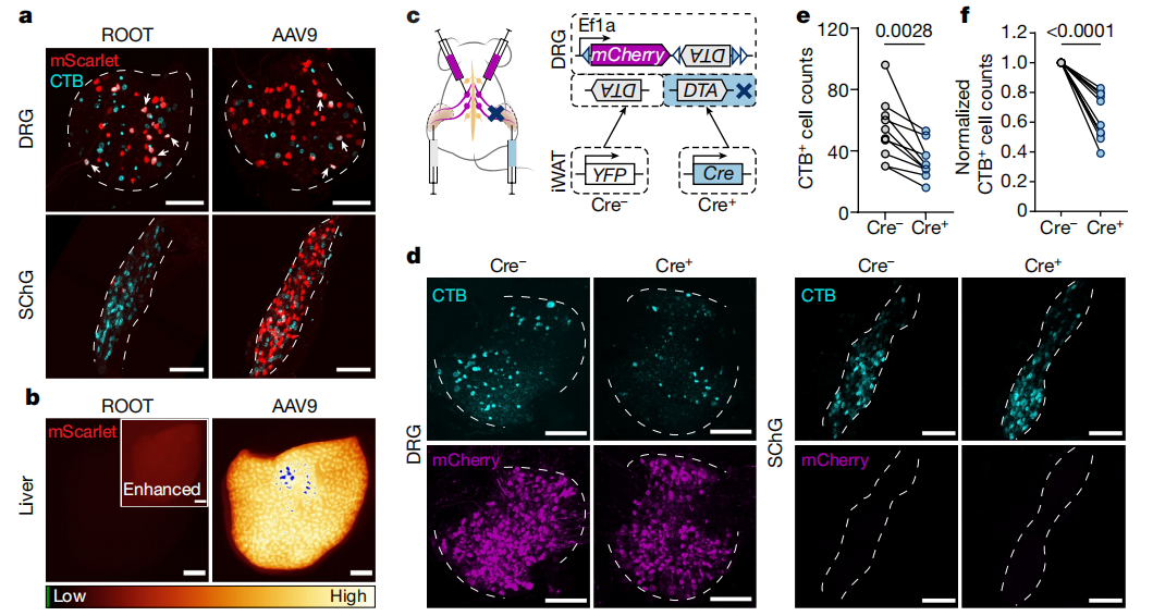
To further investigate the specific function of DRG→iWAT projections, researchers developed the ROOT retrograde tracing technology based on AAV9. The ROOT capsid library was generated by inserting a random heptamer between positions 588 and 589 of Gibson-assembled AAV9 using NNK degenerate primers (Integrated DNA Technologies). This modification significantly enhanced retrograde labeling efficiency, allowing for specific tracing of DRG neurons projecting to adipose tissue while minimizing off-target labeling in the sympathetic chain ganglia (SChG), contralateral DRGs, and liver (Figure 2a-b). Using the ROOT technology, researchers injected a Cre-dependent diphtheria toxin A subunit (DTA) construct (mCherry-flex-DTA) bilaterally into DRGs and unilaterally injected ROOT expressing either Cre or YFP into iWAT. The DTA gene was introduced into cells to selectively ablate sensory neurons. Results showed that, compared to controls, the number of DRG neurons innervating iWAT was reduced by approximately 40%, while the number of SChG neurons innervating iWAT remained unchanged (Figure 2c-f). Thus, by combining retrograde AAV (AAV-ROOT) with a Cre-dependent DTA system, researchers successfully achieved specific ablation of sensory neurons innervating adipose tissue.
Figure 2: Specific Labeling of Fat-Sensing Neurons
Ablation of Sensory Neurons Upregulates Thermogenesis-Related Gene Expression in iWAT
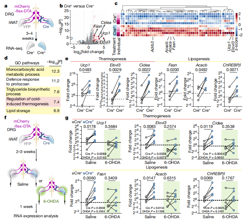
Next, researchers injected mCherry-flex-DTA bilaterally into DRGs and ROOT expressing either Cre or YFP unilaterally into iWAT. After 3–4 weeks of viral expression, they performed RNA sequencing (RNA-seq) analysis on adipose tissue from the same animal, comparing Cre⁺ (ablated side) and Cre⁻ (control side) iWAT. The results showed that the specific ablation of sensory neurons led to a significant upregulation of genes related to lipogenesis and thermogenesis, including Ucp1, Elovl3, and Cidea (Figure 3a-e). Since lipogenesis and thermogenesis are regulated by sympathetic innervation, the researchers further examined whether the sensory neuron-induced transcriptional upregulation depended on sympathetic input. In iWAT with unilateral sensory neuron ablation, they selectively destroyed sympathetic nerves using the catecholaminergic neurotoxin 6-hydroxydopamine (6-OHDA). This intervention eliminated the previously observed upregulation of thermogenesis-related genes (Figure 3f-g). These findings indicate that gene expression changes induced by sensory neuron ablation depend on an intact sympathetic nervous system. Sensory neurons regulate adipose function by inhibiting sympathetic activity.
Figure 3: Specific Loss of Sensory Neurons Upregulates Thermogenesis-Related Gene Transcription
Sensory Neuron Ablation Alters iWAT Morphology and Physiological Function
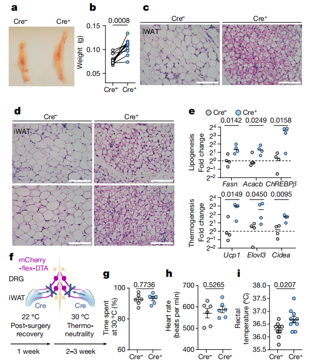
Finally, researchers investigated how sensory neuron ablation-induced gene changes affect adipose tissue function. They observed that, compared to the Cre⁻ (control side), the Cre⁺ (ablated side) iWAT exhibited:Significantly increased fat mass,Elevated phosphorylation levels of hormone-sensitive lipase (HSL),Enrichment of beige adipocytes,Significantly upregulated transcription of thermogenesis-related genes (Figure 4a-e). To further validate these findings, researchers ablated sensory innervation in bilateral iWAT and housed the mice in a thermoneutral (30°C) environment to eliminate other thermoregulatory influences. They found that mice exhibited an increase in core body temperature, while body weight, food intake, temperature sensitivity, and global sympathetic tone remained unchanged (Figure 4f-i).
Figure 4: Specific Loss of Sensory Neurons Alters iWAT Morphology and Physiological Function
Conclusion
This study revealed that DRG neurons innervate iWAT and regulate its gene transcription, thereby influencing thermogenesis. This regulatory process depends on sympathetic nervous system activity, uncovering a novel neural mechanism underlying fat-to-brain communication mediated by the DRG. The findings propose a new mechanism by which sensory neurons modulate fat metabolism and thermoregulation via sympathetic nervous system regulation. References: Wang Y, Leung VH, Zhang Y, Nudell VS, Loud M, Servin-Vences MR, Yang D, Wang K, Moya-Garzon MD, Li VL, Long JZ, Patapoutian A, Ye L. The role of somatosensory innervation of adipose tissues. Nature. 2022 Sep;609(7927):569-574. doi: 10.1038/s41586-022-05137-7. Epub 2022 Aug 31. PMID: 36045288; PMCID: PMC9477745.
Contact Us
Brain Case can provide customers with a full range of vector construction, virus packaging and stable cell line construction services. If you are interested in customized services, please contact bd@ebraincase.com for details or to place an order.