Autism Spectrum Disorder (ASD): Historical Origins, Etiology Exploration, and New Dawn in Treatment
Release time:2025-02-28 11:26:46
Autism Spectrum Disorder (ASD) is a neurodevelopmental disorder characterized by social communication difficulties, repetitive behaviors, and restricted interests. Since it was first described in the 1940s, the understanding of ASD has shifted from the “psychological trauma hypothesis” to the “neurobiological heterogeneity” model. In recent years, breakthroughs in genomics, brain imaging technologies, and artificial intelligence have propelled ASD research into the era of precision medicine. This article combines the latest literature to systematically review the scientific progression of ASD from historical evolution, etiology hypotheses, research advancements, and treatment prospects.
The research history of ASD has transitioned from psychological attributions to biological mechanisms. In 1943, Leo Kanner first described autism and proposed the “refrigerator mother” theory, attributing the cause to the mother’s emotional coldness, a theory that dominated clinical practice for a long time. In the 1960s, Bernard Rimland proposed a biological basis for ASD, leading to the development of behavioral interventions, such as ABA therapy. In recent years, MRI-based multimodal diagnostic technologies have significantly improved the early identification of ASD. A systematic review by Schielen et al. (2024) indicated that MRI can detect abnormalities in the cerebellum, amygdala, and prefrontal cortex volumes in ASD patients, with sensitivities and specificities of 82% and 76%, respectively. These structural abnormalities, along with the DSM-5’s dimensional definition (2013), support the neurobiological heterogeneity hypothesis of ASD.
The etiology of ASD is complex, involving the interaction of genes, environment, and brain networks. In terms of genetic factors, rare mutations and syndromic ASD (such as SHANK3, NLGN3/4X mutations) and chromosomal abnormalities (e.g., 15q11-13 duplication, 16p11.2 deletion) are important risk factors. Genome-wide association studies (GWAS) have also identified hundreds of common risk loci (e.g., CDH9, CNTNAP2), explaining about 20% of the genetic risk. Environmental factors play a crucial role in the onset of ASD, with maternal immune activation (MIA) caused by prenatal infections, as well as metabolic and nutritional issues such as folate deficiency and vitamin D insufficiency, potentially increasing ASD risk through epigenetic mechanisms (e.g., DNA methylation). In terms of neurobiological mechanisms, dysregulation of synaptic plasticity (e.g., SHANK3 deletion leading to impaired glutamatergic synaptic transmission) and brain network connectivity imbalance (e.g., local overconnectivity and weakened long-range connectivity) are core features of ASD. Wang et al. (2024) developed an ASD prediction model based on a graph neural network (Residual Graph Transformer), which integrated multi-center MRI and clinical data, achieving a prediction accuracy of 92.3% (AUC=0.94), significantly outperforming traditional machine learning methods. This model can identify key brain regions (e.g., the insula and cingulate gyrus), providing targets for individualized interventions.
In recent years, significant breakthroughs have been made in ASD research, including gene editing, organoid models, the gut-brain axis, and neural modulation. CRISPR screening technology has revealed the critical role of ASD risk genes (e.g., CHD8) in neurogenesis, while brain organoids derived from patient iPSCs simulate early brain development processes, revealing ASD-related defects in cortical neuron migration and GABAergic inhibition. Research on the gut-brain axis suggests that ASD children exhibit gut microbiota imbalances (e.g., reduced Prevotella and increased Clostridia), which may influence behavior through metabolic products (e.g., 4EPS). Clinical trials on fecal microbiota transplantation (FMT) show improvements in ASD children’s social behaviors and gastrointestinal symptoms. In terms of neural modulation and targeted therapies, novel treatments such as oxytocin nasal spray and mTOR inhibitors (e.g., everolimus) are showing promise in clinical trials. Tseng et al. (2024) found that elevated TSPO expression in female ASD patients was associated with astrocyte activation, suggesting that anti-inflammatory drugs (e.g., minocycline) may be effective for specific subgroups. CRISPR editing combined with AAV vector delivery technology can repair synaptic defects in SHANK3 knockout models (e.g., restoring 30% dendritic spine density). Brain Case Biotech offers gene editing based on CRISPR technology combined with AAV vector delivery services, assisting in the development of drugs for neurological diseases. 👇👇 https://www.ebraincase.com/products-service/popular-applications/gene-regulation/234.html
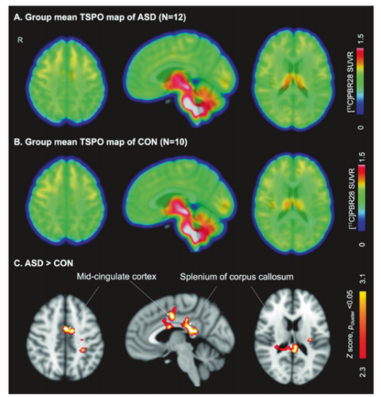
Figure 1. Compared with age and TSPO genotype-matched control groups (CON), ASD adult females exhibit increased [11C]PBR28 SUVR levels. Group averages of [11C]PBR28 SUVR for ASD females (A) and control females (B). (C) After controlling for age and TSPO genotype, voxel-based statistical comparison of [11C]PBR28 SUVR between the two groups shows significantly elevated TSPO levels in the mid-cingulate cortex and the splenium of the corpus callosum in the ASD group (N = 12) compared to the control group (N = 10), relative to the whole-brain average (Z > 2.3, pcluster< 0.05). TSPO: Translocator protein, ASD: Autism Spectrum Disorder, CON: Control group, SUVR: Standardized Uptake Value Ratio, N: Sample size.
The future development of neuroscience will rely on cutting-edge tools such as multimodal brain imaging, artificial intelligence, and big data. Duan et al. (2024) conducted a high-resolution fMRI study with 166 ASD toddlers and 109 typical development (TD) toddlers, totaling 372 scans. The study found that compared to TD toddlers, ASD toddlers exhibited increased volume or cortical thickness in the temporal lobe and fusiform gyrus, while showing decreased volume or cortical thickness in the lower frontal lobe and midline structures, revealing abnormal early brain volume growth in ASD infants. Fiber-optic spectrometry has enabled real-time monitoring of specific neuron populations in freely moving animals, elucidating the neural coding of social behavior. Deep learning algorithms analyze eye movements and hand gestures through video, achieving early ASD screening with accuracy above 90%, while integrated models of genomics, brain connectomics, and clinical data offer new approaches to personalized treatment and precision medicine. Moreover, the neurodiversity movement advocates for respecting the cognitive differences of ASD individuals, promoting inclusive education and workplace policies, and improving patients’ quality of life through digital therapies (e.g., VR social training) and community support.
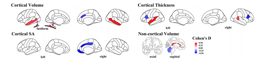
Figure 2. Significant differences between ASD and TD toddlers in cortical volume, non-cortical volume, cortical thickness, and cortical surface area. Colors represent the corresponding effect sizes (Cohen’s D), with red regions indicating significantly increased values in ASD compared to TD, and blue regions indicating significantly decreased values in ASD compared to TD. The deeper the color, the greater the difference between ASD and TD.
From the “refrigerator mother” theory to the gene-brain-environment interaction model, ASD research has undergone 80 years of groundbreaking development. Revolutionary tools in neuroscience, such as organoids and optogenetics, are gradually unveiling its etiological black box, while the integration of precision medicine and artificial intelligence brings a new dawn for individualized treatment. However, the extreme heterogeneity of ASD requires us to abandon the “one-size-fits-all” approach and instead adopt multi-level intervention strategies—from molecular repair and brain network remodeling to microbiome regulation and social support. As stated in the 2023 Science Outlook: “Understanding ASD is not only about decoding the brain, but also reshaping our cognition of human neurodiversity.”
Ref: [1]. Schielen, S. J. C., Pilmeyer, J., Aldenkamp, A. P., & Zinger, S. (2024). The diagnosis of ASD with MRI: a systematic review and meta-analysis. Translational psychiatry, 14(1), 318. https://doi.org/10.1038/s41398-024-03024-5 [2]. Wang, Y., Long, H., Bo, T., & Zheng, J. (2024). Residual graph transformer for autism spectrum disorder prediction. Computer methods and programs in biomedicine, 247, 108065.https://doi.org/10.1016/j.cmpb.2024.108065 [3]. Tseng, C. J., Canales, C., Marcus, R. E., Parmar, A. J., Hightower, B. G., Mullett, J. E., Makary, M. M., Tassone, A. U., Saro, H. K., Townsend, P. H., Birtwell, K., Nowinski, L., Thom, R. P., Palumbo, M. L., Keary, C., Catana, C., McDougle, C. J., Hooker, J. M., & Zürcher, N. R. (2024). In vivo translocator protein in females with autism spectrum disorder: a pilot study. Neuropsychopharmacology : official publication of the American College of Neuropsychopharmacology, 49(7), 1193–1201. https://doi.org/10.1038/s41386-024-01859-6 [4]. Duan, K., Eyler, L., Pierce, K., Lombardo, M. V., Datko, M., Hagler, D. J., Taluja, V., Zahiri, J., Campbell, K., Barnes, C. C., Arias, S., Nalabolu, S., Troxel, J., Ji, P., & Courchesne, E. (2024). Differences in regional brain structure in toddlers with autism are related to future language outcomes. Nature communications, 15(1), 5075. https://doi.org/10.1038/s41467-024-48952-4
Contact Us
Brain Case can provide customers with a full range of vector construction, virus packaging and stable cell line construction services. If you are interested in customized services, please contact bd@ebraincase.com for details or to place an order.