E-mail:BD@ebraincase.com
Tel:+8618971215294
English 中文版
Virus Vector - Viral Vector Production - BrainCaseVirus Vector - Viral Vector Production - BrainCase
  • Home
  • Virus product library
    CRISPRRNAiHSV-helperRV-helperNeurophilic virusCalcium SensorsOptogenetics activationOptogenetics inhibitionChemical geneticsSparse labelingFluorescent proteinBiosensorsRecombinaseApoptosis & AutophagyDisease ModelNeurotoxicityOther
  • Products & Service

    Product Center

    Virus

    VSV-circuit research-vaccine and gene therapy research-BrainCase
    Retrovirus-RCAS-TVA-BrainCase
    Lentivirus Vector - Lentivirus Production - BrainCase
    Rabies Virus Vector - RBV Vector - BrainCase
    Herpes simplex virus-Oncolytic and anterograde tracing-Brain Case
    PRV-retrograd multisynaptic-Peripheral-Btain Case
    AAV-gene therapy vectors-BrainCase

    Animal Model

    Neurological Disease Models-BrainCase
    Tumor animal models-anti-tumor-BrainCase
    Digestive System Disease Animal Model-Brain Case
    Cardiovascular System Disease Animal Models-Brain Case

    Plasmid Construction

    Bac
    Library Construction
    Plasmid design and construction

    Popular Applications

    Gene Regulation

    Gene Overexpression-Brain Case
    RNA interference(RNAi)-siRNA-Brain Case
    Gene Editing - CRSIPR cloning - BrainCase

    Neural Circuit Function Research

    Optogenetics - BrainCase
    Chemical genetics-DREADDs-Brain Case
    Calcium signal recording-Gels- Brain Case
    GRAB Neurotransmitter Fluorescent Probe- Brain Case
    Functional Magnetic Resonance Imaging Technology- Brain Case

    Research on the structure of neural circuits

    Direct Input and Output-viral vectors- Brain Case
    Anterograde Mono-synaptic Tracing -HSV- Brain Case
    Antrograde Muti-synaptic Tracing-HSV & VSV-Brain Case
    Retrograde Mono-synaptic Tracing-Rabies Virus-Brain Case
    Retrograde Muti-synaptic Tracing-PRV-Brain Case

    Featured Services

    Nervous System Disease Drug Effect

    Alzheimer's disease-AD-Brain Case
    Depression-mental disorders-Brain Case
    Parkinson's disease--PD-Brain Case
    Epilepsy-an ancient neurological disorder-Brain Case

    AAV Serotype Screening

    AAV Serotypes screening-gene therapy-Brain Case

    Tumorigenicity Test

    Tumorigenicity Testing-Evaluation of tumor models-Brain Case

    Efficacy of Oncolytic Virus

    Oncolytic virus-for cancer therapy-Brain Case
    Herpes Virus Vector-anti-tumor- BrainCase
    Vesicular stomatitis virus-killing tumor cells-Brain Case
  • News
    Corporate News New Product Launch Media Activity Investor News
  • Support
    Literature interpretation Customer article Video Zone FAQs
  • About Us
    Virus product library Products & Service News Support About Us Contact
  • Contact
    Contact Us Join us
  • 中文
    English 中文版
  • Home
  • Support
  • Literature interpretation
  • Support
  • Literature interpretation
  • Customer article
  • Video Zone
  • FAQs

Cell︱Precise Pain Control: HCAD System Revolutionizes Peripheral Chemogenetics

Release time:2025-03-05 10:20:21
Pain serves as a protective mechanism for our body, but chronic pain severely affects the quality of life for many individuals. Traditional painkillers often act systemically, including in the brain, which may lead to various side effects. Therefore, scientists have been seeking more precise methods for pain control.

On December 3, 2024, Professor Bryan L. Roth’s team from the University of North Carolina published a research paper in Cell titled "Structure-guided design of a peripherally restricted chemogenetic system."
The team developed the HCAD system, a DREADD-based chemogenetic tool utilizing the hydroxycarboxylic acid receptor 2 (HCA2).

By using a specific chemical activator, FCH-2296413, the HCAD system can precisely target peripheral nerves, particularly nociceptors in the dorsal root ganglia (DRG), to alleviate pain. The system is designed to prevent drugs from crossing the blood-brain barrier, thereby achieving precise control over peripheral tissues.


DREADD Based on Peripheral HCA2 Receptor

The HCA2 receptor was chosen as the template for the DREADD system because its expression is low in the brain and other neural tissues, while it is more abundantly expressed in innate immune cells, which are primarily located in barrier tissues. Due to the low expression of HCA2 in neurons, drugs targeting HCA2 can minimize potential effects on brain neurons, thereby reducing side effects.

MK-0354, a Gi-biased agonist of HCA2, was previously developed but did not progress to Phase II clinical trials due to its poor efficacy. However, such low-efficacy or inactive ligands are well-suited as potential chemogenetic activators in the DREADD system.

To optimize this approach, researchers mutated the HCA2 receptor to create HCAD and successfully screened a more potent and selective chemical activator, FCH-2296413.

Figure 1 Schematic Diagram of the HCAD System Principle


Validation of HCAD Peripheral Nerve Modulation Function

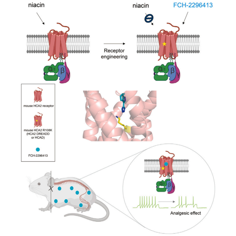
Most acute, perioperative, and chronic pain is driven by peripheral primary afferent neurons in the DRG. Therefore, reducing DRG neuron activity, particularly nociceptor activity, is a key strategy for developing advanced pain treatments.

To verify the in vivo functionality of the HCAD DREADD system, researchers developed an adeno-associated virus (AAV) to infect DRG nociceptors and express mHCAD-mCitrine in a Cre recombinase-dependent manner (PHP.S-CAG-FLEx-mHCAD-mCitrine).
In newborn (P2) Avil-Cre and TRPV1-Cre mice, AAV was administered via intraperitoneal injection, while in adult mice, it was delivered intrathecally to label DRG neurons and DRG nociceptors, respectively. Following viral injection, mHCAD-mCitrine was successfully expressed in Cre+ DRG neurons (Figure 2A).

At the cellular level, whole-cell patch-clamp recording was performed on isolated DRG neurons to assess the function of mHCAD-mCitrine. In DRG neurons, Gi signaling typically inhibits voltage-gated calcium channels (VGCCs), thereby reducing neurotransmitter release and suppressing the transmission of somatosensory information—including pain signals—from the peripheral nervous system (PNS) to the spinal cord and brain.

In Avil-Cre mice, FCH-2296413 reduced voltage-activated calcium current amplitudes in HCAD-mCitrine+ neurons in a dose-dependent manner. At a concentration of 100 nM, FCH-2296413 showed no inhibitory effect, whereas at 1 μM and 10 μM, it significantly inhibited VGCC activity (Figures 2B and 2C). Furthermore, FCH-2296413 slowed calcium current kinetics after administration (Figures 2B and 2D).

These inhibitory effects closely resemble those produced by opioid analgesics acting on endogenous Gi-GPCRs in DRG neurons. Notably, in mHCAD-mCitrine-negative (control) neurons, FCH-2296413 had no effect on VGCC activity.


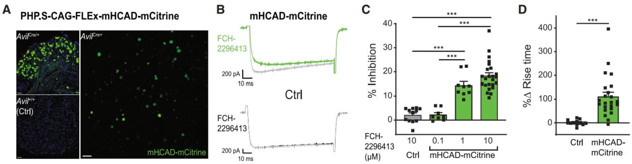
Figure 2 Whole-Cell Patch-Clamp Recording of Voltage-Activated Calcium Currents


Another typical effect of Gi signaling is membrane hyperpolarization, which leads to reduced excitability and altered somatosensory encoding. The study found that FCH-2296413 significantly decreased the resting membrane potential (RMP) of mHCAD-mCitrine+ DRG neurons (Figures 3E and 3F) and reduced action potentials (APs) (Figures 3E and 3G).

These effects were reversible, as RMP and APs returned to baseline levels after washing out FCH-2296413 (Figures 3F and 3G). Additionally, no such changes were observed in mHCAD-mCitrine-negative control neurons (Figures 3F and 3G).


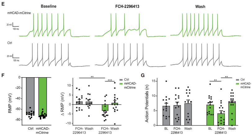
Figure 3 Effects of the mHCAD System on Resting Membrane Potential and Action Potentials


Previous reports have suggested that hM4Di expression can alter the function of ion channels in DRG neurons even in the absence of an agonist. Therefore, to further characterize mHCAD function and its potential advantages in studying peripheral neurons, researchers examined whether mHCAD expression affected ion currents involved in AP formation in the absence of FCH-2296413.

The results showed no significant differences in AP waveform threshold, overshoot (membrane potential exceeding the normal resting potential at the AP peak), or duration (Figures 4H-4M, 4I-4K). This indicates that voltage-gated sodium channels and voltage-gated potassium channels responsible for AP initiation, rise, and fall phases were not affected by mHCAD expression.

However, mHCAD expression slightly increased the amplitude of afterhyperpolarization (AHP) without altering its decay time (Figures 4L and 4M). Most importantly, there was no difference in excitability between mHCAD-mCitrine+ cells and non-expressing cells (Figure 4N). Additionally, unlike hM4Di, mHCAD expression did not induce any detectable gene expression changes (Figure 4O).

In summary,
mHCAD expression had minimal impact on DRG neuron function in the absence of FCH-2296413, whereas FCH-2296413 activation of mHCAD influenced classical Gi signaling pathways associated with reduced excitability and neurotransmitter release in DRG neurons.


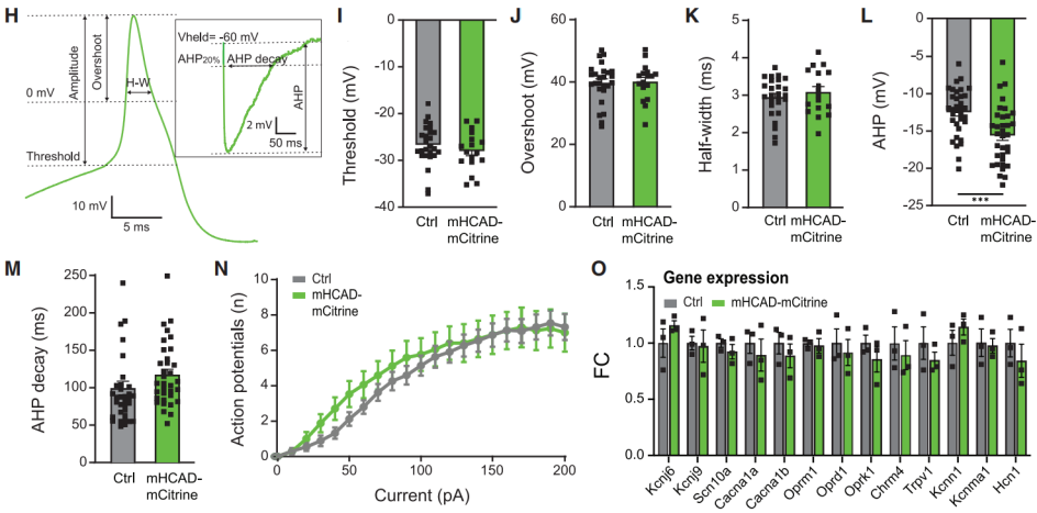
Figure 4 Effects of mHCAD on Currents Involved in Action Potential Formation


HCAD Activation in DRG Nociceptors Alleviates Pain

Researchers evaluated the effects of FCH-2296413 (10 mg/kg, subcutaneous injection) on pain perception in Trpv1-Cre mice that received an intrathecal injection of either PHP.S-CAG-FLEx-mHCAD-mCitrine or the control virus PHP.S-CAG-FLEx-tdTomato (Ctrl) (Figures 5A and 5B).

In the hot plate test for acute thermal pain (Figure 5C), FCH-2296413 significantly reduced the duration and frequency of forepaw licking, hindpaw licking, and jumping in mice expressing mHCAD in DRG nociceptors. It also increased the latency to the first nocifensive behavior, suggesting an analgesic effect mediated by mHCAD.

Furthermore, in the Freund’s adjuvant (CFA) model of tissue injury and chronic inflammatory pain (Figure 5D), FCH-2296413 alleviated mechanical and thermal hypersensitivity in mHCAD-expressing mice, but not in control mice, confirming the therapeutic anti-hyperalgesic properties of FCH-2296413 and mHCAD.

Overall, these findings demonstrate that in mice expressing mHCAD in DRG nociceptors, FCH-2296413 exerts analgesic effects on both physiological and pathological pain conditions.


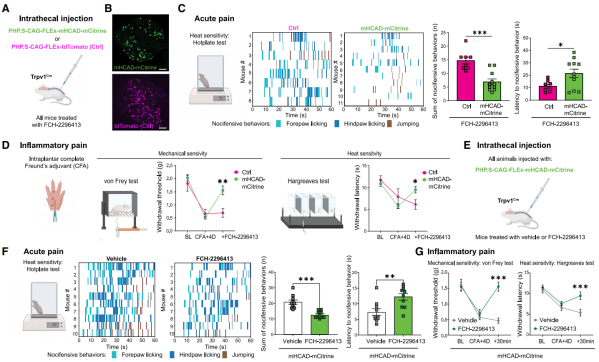
Figure 5 Peripheral Chemogenetic Analgesic Effects of mHCAD
 

Previous studies have reported that other chemogenetic drugs (such as CNO) can alter locomotor behavior in mice, potentially confounding interpretations of various behaviors, including nocifensive responses. To address this, researchers examined the effects of FCH-2296413 on various behaviors in wild-type (WT) mice that do not express mHCAD (Figures 6H-6P).

The results showed that FCH-2296413 had no effect on mechanical thresholds in WT mice and did not induce changes in nocifensive behaviors in the tail immersion test (Figure 6I), hot plate test (Figures 6J and 6K), or Hargreaves test.

Furthermore, researchers evaluated the effects of FCH-2296413 on locomotion and anxiety-like behavior in the open field test and motor coordination in the rotarod test. In these experiments,
FCH-2296413 did not affect the behavior of WT mice.

These findings indicate that the peripheral DREADD system proposed in this study can specifically modulate DRG nociceptor function and alleviate pain. This system holds potential for mechanistic studies of peripheral neuron function and the treatment of diseases affecting the PNS.


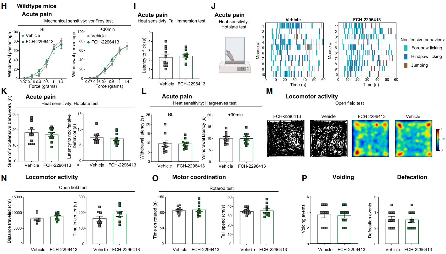
Figure 6 FCH-2296413 Does Not Affect Behavior in WT Mice


Potential Applications

This study provides a novel tool for researching and treating pain and other peripheral nervous system-related diseases, while avoiding the central nervous system side effects associated with traditional drugs. The successful development and validation of the HCAD system offer broad application prospects in neuroscience research and clinical therapy, especially in scenarios requiring precise neural activity control without affecting brain function.
 

Contact Us

Brain Case can provide customers with a full range of vector construction, virus packaging and stable cell line construction services. If you are interested in customized services, please contact bd@ebraincase.com for details or to place an order.


💥Holiday

Special

Offers

Click to fill in the requirements and submit them to us!

Service Type :

Select the service you'd like to purchase.

Order Information(Premade-AAVs)

Please provide us some information about the service you'd like to order.

Detailed requirements:

scroll

Order Information(Custom AAV/Lentivirus)

Please provide us some information about the service you'd like to order.

Gene ID or gene information:

Selection of the reporting gene:

Special Instructions:

scroll

Order Information(Others)

Please provide us some information about the service you'd like to order.

Virus name/Detailed requirements:

scroll

Related products

Breakthrough | WESTLAKE UNIVERSITY TEAM DEVELOPS ADVANCED RED GENETICALLY ENCODED CALCIUM PROBE“SOMAFRCAMPI”WITH SENSITIVITY MATCHING TO GCAMP FOR NEURONAL IMAGING IN VIVO

Breakthrough | WESTLAKE UNIVERSITY TEAM DEVELOPS ADVANCED RED GENETICALLY ENCODED CALCIUM PROBE“SOMAFRCAMPI”WITH SENSITIVITY MATCHING TO GCAMP FOR NEURONAL IMAGING IN VIVO

IF=85 Cutting-edge Review | CRISPR’s Three-Dimensional Breakthrough: A Comprehensive Overview of Genome, Epigenome, and Transcriptome Editing

IF=85 Cutting-edge Review | CRISPR’s Three-Dimensional Breakthrough: A Comprehensive Overview of Genome, Epigenome, and Transcriptome Editing

Literature Insight | Nature Methods | A “Magic Tool” for Real-Time Observation of Synaptic Structural Dynamics — SynapShot

Literature Insight | Nature Methods | A “Magic Tool” for Real-Time Observation of Synaptic Structural Dynamics — SynapShot

Breakthrough | Li Xiaojiang/Lai Liangxue team uses AAV gene therapy to treat Huntington's disease pigs

Breakthrough | Li Xiaojiang/Lai Liangxue team uses AAV gene therapy to treat Huntington's disease pigs

map
{dede:global.cfg_webname/}

Virus product library

CRISPR
RNAi
Neurophilic virus
Optogenetics activation
Biosensors

News

Corporate News
New Product Launch
Media Activity
Investor News

Support

Literature interpretation
Customer article
Video Zone
FAQs
微信

WhatsApp Business Account

Tel: +8618971215294
E-mail: BD@ebraincase.com

Address:-

Address:-

  • Copyright © 2024 Brain Case All Rights Reserved.