Cell Interpretation | CybSEP2 and NEPLDCV: Novel Genetic Tools for Monitoring and Manipulating Neuropeptide Release In Vivo
Release time:2024-12-09 15:20:40
Neuropeptides, as a class of important bioactive molecules, play indispensable roles in the nervous system. By binding to specific receptors, they participate in regulating neuron development, synaptic plasticity, and a variety of behavioral and emotional responses. Abnormal expression or dysfunction of neuropeptides is closely linked to a range of neurological diseases, including pain, depression, anxiety, schizophrenia, and neurodegenerative disorders. Traditional methods for studying neuropeptides, such as immunohistochemistry, radioimmunoassays, and molecular biology techniques, have revealed the distribution and function of neuropeptides to some extent. However, these methods often lack the ability to monitor the real-time release of neuropeptides in living animals. Therefore, researchers urgently need tools that can specifically monitor and manipulate neuropeptide release to gain a deeper understanding of the dynamic changes of neuropeptides under physiological and pathological conditions.
CELL
On July 22, 2024, Dr. Sung Han's research group at the Salk Institute for Biological Studies in the United States published a study titled "Presynaptic Sensor and Silencer of Peptidergic Transmission Reveal Neuropeptides as Primary Transmitters in Pontine Fear Circuit" online in Cell. This study introduced two novel gene-encoded tools: the LDCV sensor (CybSEP2) and the LDCV silencer (NEPLDCV), which are designed to monitor and suppress the release of neuropeptides at nerve terminals.
Design and Characterization of the LDCV Sensor
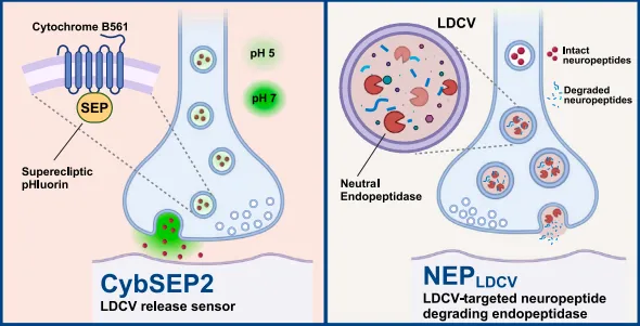
The researchers constructed the Cyb561-SEP fusion protein (CybSEP) by anchoring the SEP (Superecliptic pHluorin), a pH-sensitive version of GFP, to the luminal side of the LDCV-specific membrane protein Cytochrome b561 (Cyb561). SEP's fluorescence intensity and spectral properties change in response to environmental pH variations. It is almost completely quenched at pH 5, but shows a ~50-fold increase in green fluorescence intensity at pH 7. To determine whether CybSEP responds to pH changes with altered fluorescence, the researchers conducted experiments in PC12 cells. The results showed that CybSEP fluorescence was nearly completely lost under acidic conditions (pH 5.5), with signal recovery after NH4Cl treatment, while Cyb561-GFP fusion protein (CybGam) fluorescence remained unchanged. Treatment with 70mM KCl enhanced CybSEP fluorescence, and the fluorescence change was correlated with the frequency of electrical stimulation (10-100Hz, with the strongest response at 100Hz). EGTA treatment abolished the fluorescence changes induced by 100Hz stimulation, and TetTox inhibited fluorescence changes. Furthermore, by combining two SEPs into Cyb561 (CybSEP2), the researchers further modified CybSEP, resulting in a sensor with larger fluorescence changes and faster response times for both the rise and decay of the fluorescence following electrical stimulation.
Figure 1. Design and Characterization of CybSEP as an LDCV Sensor
Monitoring of Axonal Neuropeptide Release in Acute Brain Slices
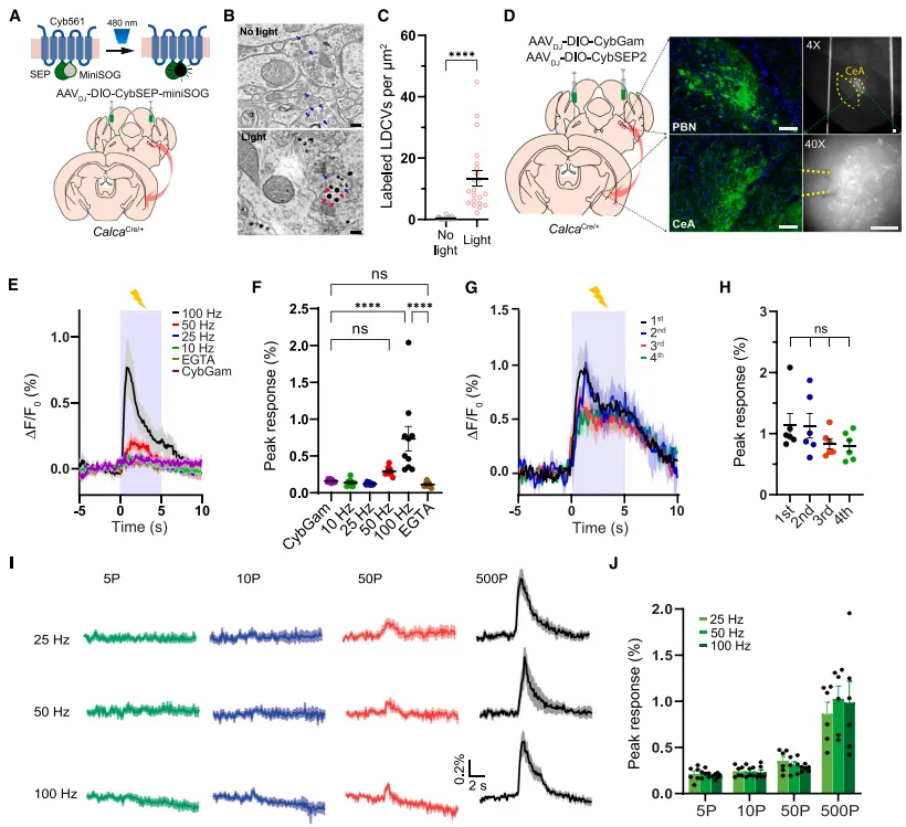
Next, the researchers used a miniaturized singlet oxygen generator (miniSOG) to observe the CybSEP localization of calcitonin gene-related peptide (CGRP)-positive neurons in the parabrachial nucleus (PBel) projecting to the central amygdala (CeAl). miniSOG, under light stimulation, catalyzes the polymerization of diaminobenzidine (DAB), which can then be observed via electron microscopy (EM). First, miniSOG was inserted between the C-terminal of SEP and the second loop of Cyb561, and packaged in an adeno-associated virus (AAVDJ-DIO-CybSEP-miniSOG). The virus was then injected into the PBel of CalcaCre/+ mice. EM imaging under light stimulation showed miniSOG labeling in LDCVs.
Additionally, by injecting AAVDJ-DIO-CybSEP2 or AAVDJ-CybGam into both sides of the PBel in CalcaCre/+ mice, the researchers observed the expression of CybSEP2 in the axons of neurons in the PBel projecting to the CeAl. Electrical stimulation showed a frequency-dependent increase in CybSEP2 fluorescence (with the strongest response at 100Hz). EGTA treatment abolished this response, and the fluorescence signal did not decay with repeated stimulation. Electrical stimulation with different pulse numbers caused an increase in CybSEP2 fluorescence, and co-expression with TetTox reduced the fluorescence change induced by 100Hz stimulation.
In conclusion, these results demonstrate that CybSEP2 can reliably detect the release of LDCVs at axon terminals in acute brain slices.
Figure 2. Imaging Neuropeptide Release at Axon Terminals of PBel→CeAl Projections in Brain Slices
Monitoring Neuropeptide Release at Axon Terminals in Freely Moving Mice
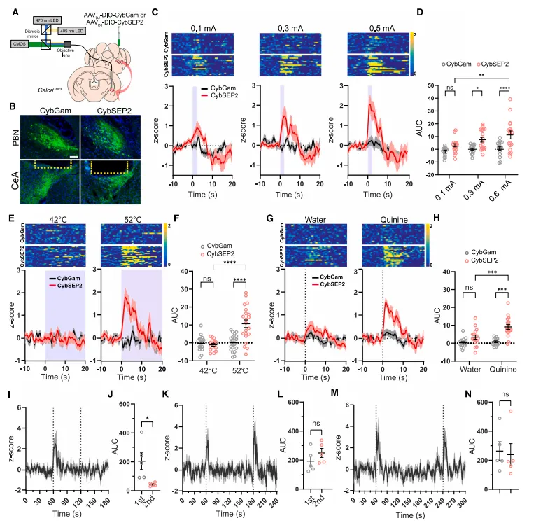
To further investigate the release of neuropeptides at CGRP PBel→CeAl axon terminals in freely moving mice under threat conditioning, the researchers injected AAVDJ-DIO-CybSEP2 or AAVDJ-DIO-CybGam into the PBel of CalcaCre/+ mice. Optical fibers were then implanted above the CeAl to monitor neuropeptide release using a CMOS fiber photometry system. The results showed that electrical stimulation at different intensities, 52°C heat stimulation, and quinine stimulation all led to an increase in fluorescence at the CGRP PBel→CeAl terminals in CybSEP2-expressing mice, whereas no fluorescence change was observed in CybGam-expressing mice.
Additionally, the researchers tested the LDCV cycling dynamics by applying two tail pinches with a 2- or 3-minute interval, which induced an increase in CybSEP2 activity, while no response was observed within a 1-minute interval.
Finally, to assess the response of CybSEP2 to optogenetic stimulation, CybSEP2 and ChrimsonR-mRuby2-ST were expressed in the PBel of CalcaCre/+ mice, and the activity of CybSEP2 in the CeAl of awake mice was measured using CMOS fiber photometry. The researchers observed that CybSEP2 fluorescence increased rapidly upon light stimulation.
In conclusion, these results demonstrate that aversive sensory stimuli can trigger the release of LDCVs at CGRP PBel→CeAl axon terminals.
Figure 3. Monitoring Neuropeptide Release at Synaptic Terminalsin Behaving Mice
Design and Characterization of the LDCV Silencer
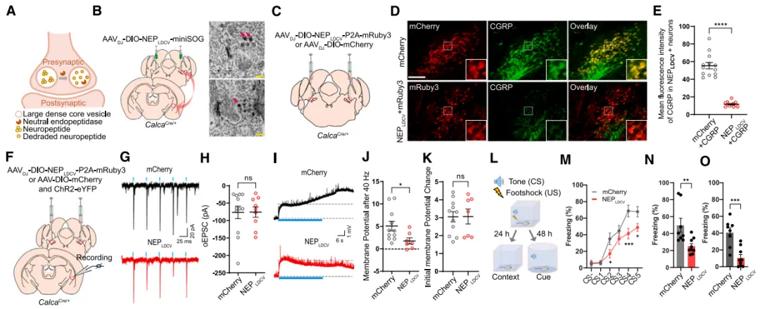
Although existing methods, such as drug blockade and gene knockout, provide valuable tools for inhibiting peptidergic signaling, there has been a lack of a universal tool that can specifically dampen peptidergic transmission in particular cell types. The researchers constructed an LDCV silencer (NEPLDCV) by specifically transporting the neuropeptide-specific enzyme, neutral endopeptidase (NEP), to the lumen of LDCVs. NEPLDCV works by cleaving hydrophobic amino acid chains, selectively inactivating a variety of neuropeptides, including but not limited to enkephalins, bradykinin, calcitonin gene-related peptide (CGRP), substance P, neurotensin, and oxytocin.
The localization of NEPLDCV in LDCVs was confirmed through miniSOG labeling, and electron microscopy (EM) imaging revealed dense labeling in the LDCVs of both cell bodies and axon terminals. Researchers injected AAVDJ-DIO-NEPLDCV-P2A-mRuby3 or AAVDJ-DIO-mCherry into both sides of the PBel in CalcaCre/+ mice. The results showed that most mCherry-expressing neurons co-localized with CGRP immunofluorescence, while neurons expressing mRuby exhibited almost no detectable CGRP immunofluorescence, indicating that NEPLDCV effectively inhibited CGRP expression.
Electrophysiological recordings showed that NEPLDCV did not affect light-evoked excitatory postsynaptic currents (oEPSCs) but significantly weakened oEPSPs induced by 40Hz stimulation, indicating that NEPLDCV selectively dampened neuropeptide transmission without altering glutamatergic transmission.
Figure 4. NEPLDCV Reduces Neuropeptide Release and Weakens Threat Learning
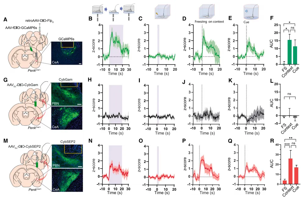
Monitoring Endogenous Opioid Release in Behaving Mice
After confirming that CybSEP2 can be used to monitor the release of CGRP from PBel→CeAl LDCVs in behaving mice, the researchers further applied CybSEP2 to study the peptidergic pathways of enkephalins, testing its multifunctionality. Enkephalins are endogenous opioid peptides that are widely distributed throughout the body and exert diverse biological regulatory effects by binding to different types of opioid receptors.
The researchers injected AAVDJ-DIO-CybGam or AAVDJ-DIO-CybSEP2 into the CeAl of PenkCre/+ mice and implanted optical fibers in the PBL. In elevated platform and classical auditory threat conditioning tests, a significant increase in fluorescence intensity was observed in the CybSEP2-expressing group, while no fluorescence changes were detected in the control CybGam-expressing group.
Figure 5. Monitoring the Release of Endogenous Opioids at ENK CeA→PBL Terminals in Behaving Mice
Conclusion
In conclusion, the researchers report two genetic-encoded tools for studying peptidergic transmission in living mice: one is the genetically encoded LDCV sensor CybSEP2, which detects presynaptic neuropeptide release, and the other is the genetically encoded LDCV silencer NEPLDCV, which specifically degrades neuropeptides within LDCVs. The development of these presynaptic sensors and silencers for peptidergic transmission provides new perspectives and methodologies for neuroscience research, aiding in a deeper understanding of the role of neuropeptides in the brain and their changes under disease conditions. Further applications of these tools could reveal more about the mechanisms by which neuropeptides contribute to brain function and disease, providing scientific basis for developing new therapeutic strategies.
References
Kim DI, Park S, Park S, Ye M, Chen JY, Kang SJ, Jhang J, Hunker AC, Zweifel LS, Caron KM, Vaughan JM, Saghatelian A, Palmiter RD, Han S. Presynaptic sensor and silencer of peptidergic transmission reveal neuropeptides as primary transmitters in pontine fear circuit. Cell. 2024 Sep 5;187(18):5102-5117.e16. doi: 10.1016/j.cell.2024.06.035. Epub 2024 Jul 22. PMID: 39043179; PMCID: PMC11380597.
Brain Case offers various virus packaging customization services. For more information, please contact bd@ebraincase.com.
Service Type :
Select the service you'd like to purchase.
Order Information(Premade-AAVs)
Please provide us some information about the service you'd like to order.
Order Information(Custom AAV/Lentivirus)
Please provide us some information about the service you'd like to order.
Order Information(Others)
Please provide us some information about the service you'd like to order.