- E-mail:BD@ebraincase.com
- Tel:+8618971215294
"Sneeze!" It's the pollen season again, and many friends will face the trouble of pollen allergies. After taking allergy medicine, allergy symptoms are relieved, but you may feel drowsy. Why is this? It turns out that anti-allergy medications contain antihistamines. When antihistamines enter the brain through the blood-brain barrier and block central histamine receptors, they cause drowsiness. So, what is histamine? Why does blocking the central histamine receptor cause drowsiness?
Histamine (HA) is an important monoamine signaling molecule that exists in the central nervous system and peripheral tissues and is widely involved in the regulation of immunity, digestion and nerve signals. Antihistamines are often used to relieve allergic reactions, but a major side effect of antihistamines is drowsiness, which has caused scientists to pay attention to the function of histamine in the central system.
In the brain, the cell bodies of histaminergic neurons are mainly distributed in the tuberomammillary nucleus (TMN) of the hypothalamus, and their nerve fibers project widely throughout the brain. Histaminergic signaling is involved in regulating important physiological processes such as sleep and awakening, learning and memory, and food intake, but its molecular regulatory mechanism remains unclear. Therefore, in order to reveal the molecular regulation mechanism of histamine on physiological processes such as sleep and wakefulness, we urgently need to develop sensitive, specific and effective tools that can monitor histamine with high spatial and temporal resolution.
On March 15, 2023, Li Yulong Laboratory of Peking University published a research paper titled Genetically encoded sensors for measuring histamine release both in vitro and in vivo online in Neuron magazine, reporting the development of a new genetically encoded histamine probe GRABHA. , and the study of dynamic regulation of histamine during sleep and awakening by combining new tools.
In this work, Li Yulong's laboratory first flexibly used the GRAB strategy (GPCR-Activation Based Sensor) to graft the cyclically rearranged green fluorescent protein cpEGFP into the third intracellular loop of four human histamine GPCR receptors and found that The H4R-based probe showed better cell membrane localization properties than other receptors. Through a series of subsequent optimizations, the H4R-based histamine fluorescent probe GRABHA1h (referred to as HA1h) was obtained. The HA1h probe expressed in HEK293T cells had ~370% fluorescence signal response (ΔF/F0) and ~17 nM affinity (EC50) to histamine (Figure 1). In order to further expand the detection range of histamine concentration by the probe, the authors screened histamine receptors from different species and developed the histamine fluorescent probe GRABHA1m (referred to as HA1m) based on tardigrade H1R. The HA1m probe The maximum ΔF/F0 of histamine is ~590%, EC50 ~380 nM. The HA1h and HA1m probes can respond to changes in extracellular histamine concentration on a sub-second (~0.3-0.6 s) time scale, and are not coupled to GPCR downstream intracellular signals; further research found that the HA1h and HA1m probes only respond Histamine, but does not respond to histamine precursors, histamine metabolites and other neurotransmitters, and the signal triggered by histamine can be blocked by antagonists of the corresponding receptors, indicating that the probe retains the corresponding receptors pharmacological properties.
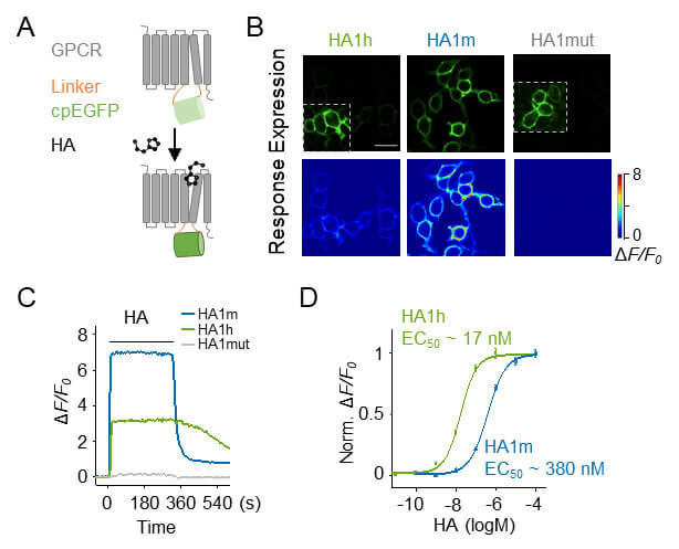
Figure 1: Characterization of the novel histamine probe. A, How the histamine probe works. B, Expression of histamine probes HA1h, HA1m and HA1mut on HEK293T cells and their response to histamine. C, Histamine probe response curve to saturating concentration of histamine. D, Signal responses of histamine probes HA1h and HA1m to different concentrations of histamine.
Are histamine probes capable of detecting the release of endogenous histamine? The authors used AAV virus to express the HA1m probe in the prefrontal cortex (PFC) of mice. After preparing acute brain slices, they successfully recorded the second-level release of histamine induced by electrical stimulation using two-photon imaging. In addition, the authors observed in real time the spread of histamine signals outward from the stimulation site. Therefore, the HA1m histamine probe enables high spatial and temporal resolution detection of endogenous histamine release.
Can histamine probes be used in living animals? Histamine is an important molecule in regulating sleep and wakefulness. The preoptic area (POA) is an important brain area involved in the regulation of sleep and wakefulness and receives dense projections from histaminergic neurons. The author first used AAV virus to express the HA1m probe in the preoptic area of mice, recorded the fluorescent signal of the probe through optical fiber, and simultaneously recorded the brain and electromyography of mice to determine the sleep state. The authors found that histamine signaling increased during the transition from rapid eye movement (REM) sleep to wakefulness and from non-rapid eye movement (NREM) sleep to wakefulness in mice, and during the transition from wakefulness to NREM sleep and NREM sleep to REM sleep. Histamine signaling decreases.
As a control, no obvious signal changes were observed using the mutation-inactivated probe, indicating the specificity of signal detection. Furthermore, the authors found that the HA1m probe signal did not change significantly in histamine synthase knockout (HDC KO) mice. In summary, the HA1m probe can achieve reliable, real-time and specific detection of dynamic changes in histamine in living mice.
Histamine neuron cell bodies are located in the tuberomammillary nucleus of the hypothalamus and send widespread projections throughout the brain. So, is the dynamic change pattern of histamine in different brain regions consistent? The authors expressed the HA1h probe in the preoptic area and prefrontal cortex of mice, and simultaneously recorded the dynamic changes of histamine in these two brain areas during sleep and awakening (Figure 2). The results showed that the signal sizes of histamine probes in the two brain regions were consistent during the same sleep phase.
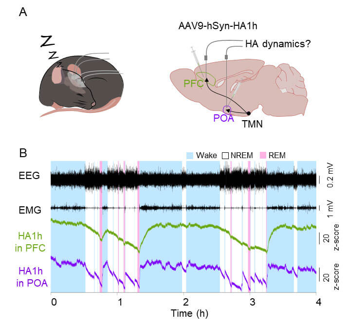
Figure 2: There are differences in the dynamic change dynamics of histamine in different nuclei during sleep phase transition.
Interestingly, during the transition from REM sleep to wakefulness, NREM sleep to wakefulness, and wakefulness to NREM sleep, the dynamics of histamine changes in the preoptic area are faster than those in the prefrontal cortex, suggesting that histamine is activated in different brain regions. There are different control modes. In conclusion, using the newly developed histamine probe, the authors found differences in histamine dynamics in different brain regions, providing important implications for studying the function of histamine.
In the central nervous system, in addition to the regulation of sleep and wakefulness studied in this work, histamine is also involved in other physiological and pathological processes, including feeding, movement, cognition, epilepsy, and migraine. In the peripheral system, histamine is also involved in processes such as allergic reactions, itching, and gastric acid secretion. The new histamine fluorescent probe will become a powerful tool to explore the molecular mechanism of histamine regulating important physiological and pathological processes.
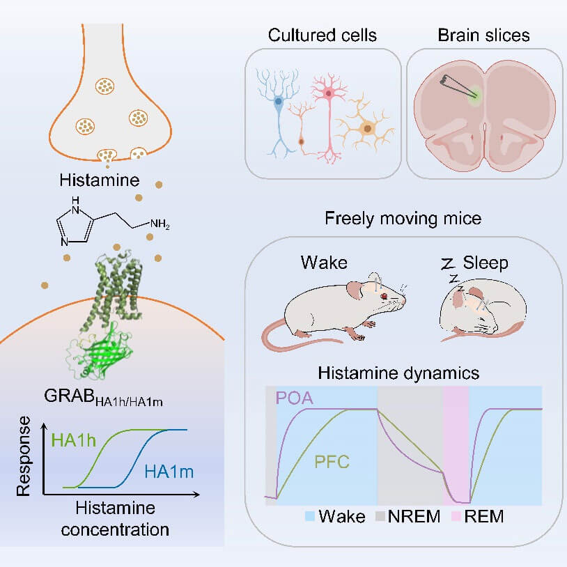
Figure 3. Working principle and in vitro and in vivo applications of the histamine fluorescent probe GRABHA.
Postdoctoral fellow Dong Hui and doctoral student Li Mengyao (graduated) from the School of Life Sciences of Peking University are the co-first authors of this article. Professor Li Yulong is the corresponding author. Laboratory members Yan Yuqi, Qian Tongrui, Lin Yunzhi, Liu Can, Li Guochuan and Wang Huan made important contributions to the article. This work was supported by the full cooperation of Professor Rob Leurs and his team Xiaoyuan Ma and Henry F. Vischer from Vrije Universiteit Amsterdam, as well as the team of Professor Chen Zhong from Zhejiang University of Traditional Chinese Medicine/Zhejiang University and the team of Professor Yang Xiangdong from Zhongshan Hospital of Fudan University. Great support. This work was supported by the National Key Laboratory of Membrane Biology of Peking University, the Peking University-Tsinghua Joint Center for Life Sciences, the National Natural Science Foundation of China, the Beijing Municipal Science and Technology Commission, the Life Medicine Summit Fund, and the Song Chenfeng and Gao Xinxin Charity Foundation. strong support.
For more details about the work in Yulong Li's laboratory, please see: http://yulonglilab.org/ In addition, Yulong Li's laboratory is looking for associate researchers, postdoctoral fellows and technicians with different disciplinary backgrounds, and the remuneration is favorable. Aspiring young people interested in brain science are welcome to join!
https://doi.org/10.1016/j.neuron.2023.02.024

The first-generation histamine H1 receptor blockers easily pass through the blood-brain barrier, and the most common side effect is drowsiness. However, histamine H3 receptor antagonists have negative feedback to increase the release of wake-related transmitters such as histamine and have been approved for the treatment of narcolepsy. Genetic and pharmacological evidence shows that the central neurotransmitter histamine has an awakening-promoting effect. However, the dynamic change and release patterns of histamine and other awakening transmitters in the brain need to be carefully analyzed. Early microdialysis methods revealed that levels of these wakefulness transmitters are high during wakefulness and decreased during sleep. However, due to the low time resolution of microdialysis methods, it is difficult for us to glimpse the details and differences in the release of these awakening transmitters. Li Yulong's team at Peking University developed a new fluorescent probe for detecting extracellular histamine through the GRAB strategy. The probe has high sensitivity and strong specificity. Using the new histamine probe, they further studied the dynamic changes of histamine in different phases of the brain in the sleep-wake-related nuclei of mice. Interestingly, there are differences in the dynamics of histamine changes in the preoptic area and prefrontal cortex of mice, suggesting that histamine may function differently in different nuclei. The article reports two histamine probes with different affinities, providing more options for detecting histamine at different concentrations. The histamine probe developed by Li Yulong's team provides a powerful tool for studying the physiological, pathological and pharmacological effects and mechanisms of histamine. The series of neurotransmitter fluorescent probes previously developed by Li Yulong's team provide strong support for our in-depth understanding of the different sleep-wake-related transmitter and modulatory functions in the brain and deciphering the mysteries of the brain.
In 1910, British scientist Dale and his colleagues reported a substance that could stimulate smooth muscle contraction and named it histamine. In the early days, histamine was thought to be mainly involved in inflammatory reactions, and antihistamine drugs were mainly used to treat allergic diseases. However, in clinical application, antihistamines are often reported to cause central nervous system-related adverse reactions such as drowsiness. Based on this clue, scientists discovered that there is a group of histaminergic neurons in the tuberomammillary nucleus (TMN) of the hypothalamus in the brain. These neurons play an important role in maintaining wakefulness. In fact, the wakefulness-promoting effect of histamine is specifically reflected in promoting brain functions such as memory and movement. Our laboratory has long been interested in the regulation of memory by histamine. A series of previous works found that histamine promotes spatial memory acquisition by increasing the excitability of glutamatergic neurons in the entorhinal cortex (Chao et al., Cerebral Cortex, 2016; Chen et al., Cerebral Cortex, 2018). How does histamine dynamically change during the wake-sleep cycle? How do different histamine levels regulate different brain functional activities? Previous research methods mainly include in-body field potential recording, calcium imaging, etc., which indirectly reflect the level of histamine release by monitoring the functional activity of histaminergic neuron cell bodies. Limited by technical means, it is difficult to clarify the release of histamine in different functional target areas and its dynamic changes. Professor Li Yulong’s team at Peking University has been committed to developing new neurotransmitter fluorescent probes based on GPCRs for many years. These new probes have been used to dynamically detect a variety of neurotransmitters in vivo, including acetylcholine, dopamine, norepinephrine, serotonin, etc. Dynamic changes. In the latest issue of Neuron, the team reported the successful development of a new histamine fluorescent probe. After thousands of screenings and optimizations, a new histamine fluorescent probe with fast response speed, high affinity, and high specificity was developed, successfully detecting dynamic changes in histamine in specific functional brain areas. More interestingly, using histamine fluorescent probes, the team found that histamine levels in the preoptic area and prefrontal cortex were both higher during wakefulness than during sleep. Changes in histamine levels in the preoptic area and prefrontal cortex show different dynamics. During the transition from sleep to awakening, histamine rises faster in the preoptic area than in the prefrontal cortex. There is no doubt that the development of new histamine probes provides a powerful tool for studying how histamine regulates different brain functional activities, and will greatly expand our understanding of the mechanism of histamine's effects. Not only that, this study found that the release of histamine in different functional brain regions has different kinetic characteristics, suggesting the heterogeneity of histamine neurons, which can provide further insights into the structure and function of histamine neurons at the single-cell level. Important clues. What needs to continue to be paid attention to is that in addition to receiving histaminergic fibers, functional brain areas such as the prefrontal cortex also receive a large number of fibers from other arousal nervous systems (such as norepinephrine, acetylcholine, etc.). How do different wake-promoting transmitters The problem of releasing and working together is a major challenge in elucidating how arousal systems work. We look forward to Professor Li Yulong's team developing technology that can simultaneously monitor the release of multiple neurotransmitters in order to comprehensively explore the release patterns of various wake-promoting neurotransmitters under different brain functional states. 2018). How does histamine dynamically change during the wake-sleep cycle? How do different histamine levels regulate different brain functional activities? Previous research methods mainly include in-body field potential recording, calcium imaging, etc., which indirectly reflect the level of histamine release by monitoring the functional activity of histaminergic neuron cell bodies. Limited by technical means, it is difficult to clarify the release of histamine in different functional target areas and its dynamic changes. Professor Li Yulong’s team at Peking University has been committed to developing new neurotransmitter fluorescent probes based on GPCRs for many years. These new probes have been used to dynamically detect a variety of neurotransmitters in vivo, including acetylcholine, dopamine, norepinephrine, serotonin, etc. Dynamic changes. In the latest issue of Neuron, the team reported the successful development of a new histamine fluorescent probe. After thousands of screenings and optimizations, a new histamine fluorescent probe with fast response speed, high affinity, and high specificity was developed, successfully detecting dynamic changes in histamine in specific functional brain areas. More interestingly, using histamine fluorescent probes, the team found that histamine levels in the preoptic area and prefrontal cortex were both higher during wakefulness than during sleep. Changes in histamine levels in the preoptic area and prefrontal cortex show different dynamics. During the transition from sleep to awakening, histamine rises faster in the preoptic area than in the prefrontal cortex. There is no doubt that the development of new histamine probes provides a powerful tool for studying how histamine regulates different brain functional activities, and will greatly expand our understanding of the mechanism of histamine's effects. Not only that, this study found that the release of histamine in different functional brain regions has different kinetic characteristics, suggesting the heterogeneity of histamine neurons, which can provide further insights into the structure and function of histamine neurons at the single-cell level. Important clues. What needs to continue to be paid attention to is that in addition to receiving histaminergic fibers, functional brain areas such as the prefrontal cortex also receive a large number of fibers from other arousal nervous systems (such as norepinephrine, acetylcholine, etc.). How do different wake-promoting transmitters The problem of releasing and working together is a major challenge in elucidating how arousal systems work. We look forward to Professor Li Yulong's team developing technology that can simultaneously monitor the release of multiple neurotransmitters in order to comprehensively explore the release patterns of various wake-promoting neurotransmitters under different brain functional states. 2018). How does histamine dynamically change during the wake-sleep cycle? How do different histamine levels regulate different brain functional activities? Previous research methods mainly include in-body field potential recording, calcium imaging, etc., which indirectly reflect the level of histamine release by monitoring the functional activity of histaminergic neuron cell bodies. Limited by technical means, it is difficult to clarify the release of histamine in different functional target areas and its dynamic changes. Professor Li Yulong’s team at Peking University has been committed to developing new neurotransmitter fluorescent probes based on GPCRs for many years. These new probes have been used to dynamically detect a variety of neurotransmitters in vivo, including acetylcholine, dopamine, norepinephrine, serotonin, etc. Dynamic changes. In the latest issue of Neuron, the team reported the successful development of a new histamine fluorescent probe. After thousands of screenings and optimizations, a new histamine fluorescent probe with fast response speed, high affinity, and high specificity was developed, successfully detecting dynamic changes in histamine in specific functional brain areas. More interestingly, using histamine fluorescent probes, the team found that histamine levels in the preoptic area and prefrontal cortex were both higher during wakefulness than during sleep. Changes in histamine levels in the preoptic area and prefrontal cortex show different dynamics. During the transition from sleep to awakening, histamine rises faster in the preoptic area than in the prefrontal cortex. There is no doubt that the development of new histamine probes provides a powerful tool for studying how histamine regulates different brain functional activities, and will greatly expand our understanding of the mechanism of histamine's effects. Not only that, this study found that the release of histamine in different functional brain regions has different kinetic characteristics, suggesting the heterogeneity of histamine neurons, which can provide further insights into the structure and function of histamine neurons at the single-cell level. Important clues. What needs to continue to be paid attention to is that in addition to receiving histaminergic fibers, functional brain areas such as the prefrontal cortex also receive a large number of fibers from other arousal nervous systems (such as norepinephrine, acetylcholine, etc.). How do different wake-promoting transmitters The problem of releasing and working together is a major challenge in elucidating how arousal systems work. We look forward to Professor Li Yulong’s team launching
Since its discovery in 1910, histamine has long been considered to function as a peripheral local hormone, involved in regulating gastrointestinal smooth muscle contraction and immune-inflammatory responses. It was not until around the 1970s that histamine was discovered in the brain and identified as a central neurotransmitter or neuromodulator. Interestingly, the central histaminergic nervous system is highly conserved among species. There are approximately 64,000 histaminergic neurons in the human brain, all of which are concentrated in a small nucleus called the tuberomammillary nucleus of the hypothalamus. However, The axons they send out innervate nearly the entire brain. Therefore, histamine is considered to play the role of a "regulator of whole brain functions" and is widely involved in the regulation of a variety of basic physiological homeostasis and advanced functions such as sleep and wakefulness, feeding rhythms, motor control, learning and memory, and emotional rewards. Four histamine receptor subtypes (H1R-H4R) have been identified successively, and they all belong to G protein-coupled receptors (GPCRs). It is worth noting that the histamine receptor family is one of the receptor families with the highest investment-output ratio in drug development at present, and histaminergic drugs have been widely used in the clinical treatment of allergies, digestive system ulcers, and vestibular diseases. In addition, studies have shown that the central histaminergic nervous system is also closely related to sleep disorders, narcolepsy-cataplexy, Tourette syndrome, multiple sclerosis, Parkinson's disease, epilepsy, and alcohol addiction. Therefore, elucidating the dynamic activity patterns of central histamine in various physiological functions and pathological changes is of great significance for in-depth understanding of the brain function regulation mechanism of the central histaminergic nervous system and the development of new histaminergic drugs. However, highly sensitive histamine probing tools with spatiotemporal resolution are still lacking. The team led by Professor Li Yulong of Peking University is one of the top international teams in developing genetically encoded fluorescent probes for detecting neurotransmitters/modulators. Its latest development of genetically encoded histamine based on G protein-coupled receptor (GPCR) activation The probe enables high spatial and temporal resolution monitoring of extracellular histamine dynamics in ex vivo brain slices and freely moving mouse brains. Their research work published in Neuron reported for the first time the dynamic activity patterns of histamine signaling in a continuous and complete sleep-wake cycle, and discovered that the histamine signaling in the key sleep-wake regulation center, the preoptic area (POA), is There was a significant increase in the transition from rapid eye movement (REM) sleep to wakefulness and the transition from non-rapid eye movement (NREM) sleep to wakefulness, and a significant decrease in the transition from wakefulness to NREM sleep and NREM sleep to REM sleep. . In particular, the team compared and analyzed the receptive histaminergic fibers that densely project and play key mediators in executive functions closely related to wakefulness, such as attention and decision-making. The histamine signal in the prefrontal cortex (PFC) and POA, which regulates the role of The awakening transition and the transition from awakening to NREM sleep show completely different dynamic characteristics, which opens up new ideas for the future in-depth analysis of the neural mechanisms and coding patterns of the interaction and coordinated integration between sleep-wake and executive functions. It is foreseeable that the successful development and continuous iteration of high spatiotemporal resolution histamine probes will provide a powerful new tool for finely characterizing the dynamic changes of central histamine in different physiological and pathological processes, allowing re-analysis in the high spatiotemporal resolution dimension. It becomes possible to understand the activity patterns of the central histaminergic nervous system, thereby injecting new vitality into a comprehensive understanding of the new functions of central and even peripheral histamine, understanding of new mechanisms for the occurrence and development of related diseases, and the development of new strategies for intervention and treatment.
If you need to reprint, please contact the editor on WeChat: brainnews_01 or email: brainnews@vip.163.com


WhatsApp Business Account
Address:-
Address:-


