- E-mail:BD@ebraincase.com
- Tel:+8618971215294
On June 13, 2023, the research team of Zhang Xu of the Guangdong Institute of Intelligence Science and Technology (referred to as: Guangdong Institute of Intelligence) published a paper titled "A direct spino- "cortical circuit bypassing the thalamus modulates nociception" paper. This study comprehensively used experimental methods such as neural circuit tracing, electron microscopy, calcium imaging, and behavioral methods to discover a new direct spinal cord-cortical pathway that conducts pain. This study provides a new way to understand brain processing. Sensory information such as pain provides new nerve conduction pathway mechanisms.

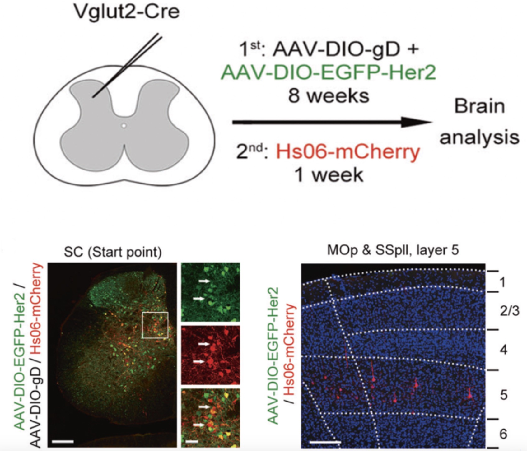
When animals experience noxious stimuli on their body surface, nociceptive signals typically travel from the spinal cord to the brain via the thalamus and then to the cortex. Here, we present a set of direct input circuits from the spinal cord to the cortex that bypass the thalamus, defining these neurons as spino-cortical recipient neurons (SCRNs). To confirm this direct spino-cortical connection, researchers utilized anterogradely trans-monosynaptic tracing using a genetically modified H129 mutant virus (named Hs06). Moreover, Hs06 could efficiently package its progenies with compensatory expression of codon-optimized glycoprotein D (cmgD) in the defined neurons, and then infect the downstream neurons trans-monosynaptically. To anterogradely trace the recipients of Vglut2-positive spinal projection neurons, Researchers injected Hs06 labeled with the red fluorescent protein (Hs06-mCherry) following the expression of adeno-associated virus (AAV) helpers AAV2/9-hSyn-DIO-EGFP-T2A-Her2CT9 (AAV-DIO-EGFP-Her2) and AAV2/9-UL26.5p-DIO-cmgD (AAV-DIO-gD) in the lumbar spinal cord of Vglut2-Cre mice. Researchers observed both EGFP/mCherry positive start neurons and mCherry-positive second-order neurons in the spinal cord (Fig. 1h). theyfound mCherry other than EGFP expression in a population of layer 5 neurons in the SSpll and Mop (Fig. 1h), indicating that Hs06-mCherry in the infected SPNs could be anterogradely transported into the cortical layer 5 neurons. As a control, Hs06 could not infect spinal neurons without expressing Her2(Supplementary information), showing the specific infection phenotype of Hs06 virus.

Fig1:Researcher injected Hs06 labeled with the red fluorescent protein (Hs06-mCherry) following the expression of adeno-associated virus (AAV) helpers AAV-DIO-EGFP-Her2 and AAV-DIO-gD in the lumbar spinal cord of Vglut2-Cre mice, then they observed both EGFP/mCherry positive start neurons and mCherry-positive second-order neurons in the spinal cord (Fig1). Furthermore, mCherry expression was found in a population of layer 5 neurons in both SSpll and MOp, instead of EGFP.
If you want to learn more excellent research in this article, please refer to the original: https://www.nature.com/articles/s41422-023-00832-0
BC-HSV-Hs06 H129-dgD-hUbC-mCherry-P2A-scHer2::gd;
BC-1245 rAAV-hSyn-DIO-EGFP-T2A-Her2CT9;
BC-1356 rAAV-UL26.5-DIO-cmgD